


Structural and Kinetic Evaluation of Phosphoramidate Inhibitors on Thermolysin
Kljajic, M., Gerber, H.-D., Heine, A., Klebe, G.To be published.
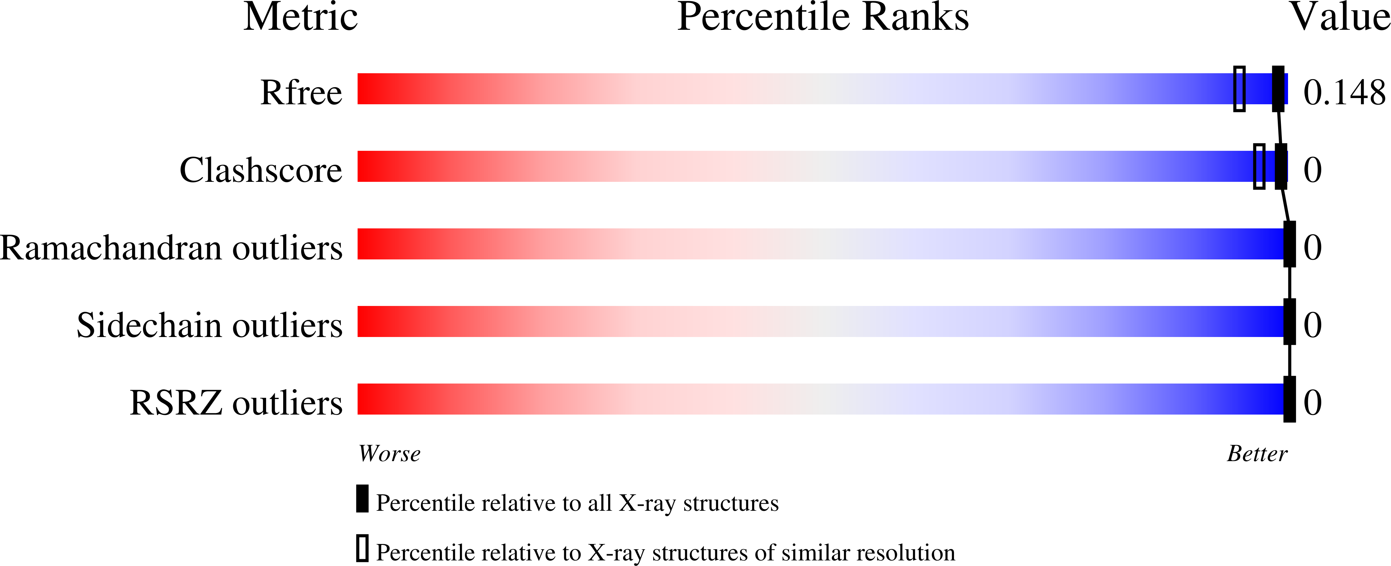
Experimental Data Snapshot
Entity ID: 1 | |||||
|---|---|---|---|---|---|
| Molecule | Chains | Sequence Length | Organism | Details | Image |
| Thermolysin | A [auth E] | 316 | Geobacillus stearothermophilus | Mutation(s): 0 Gene Names: nprS, nprM EC: 3.4.24.27 | 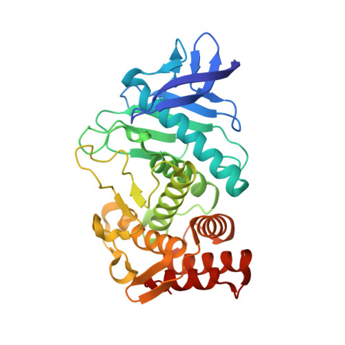 |
UniProt | |||||
Find proteins for P43133 (Geobacillus stearothermophilus) Explore P43133 Go to UniProtKB: P43133 | |||||
Entity Groups | |||||
| Sequence Clusters | 30% Identity50% Identity70% Identity90% Identity95% Identity100% Identity | ||||
| UniProt Group | P43133 | ||||
Sequence AnnotationsExpand | |||||
| |||||
| Ligands 6 Unique | |||||
|---|---|---|---|---|---|
| ID | Chains | Name / Formula / InChI Key | 2D Diagram | 3D Interactions | |
| OZH (Subject of Investigation/LOI) Query on OZH | J [auth E] | (2~{S})-4-methyl-2-[2-[[oxidanyl-[(1~{S})-2-phenyl-1-(phenylmethoxycarbonylamino)ethyl]phosphoryl]amino]ethanoylamino]pentanoic acid C24 H32 N3 O7 P YJSHTZYDPJRJOL-UNMCSNQZSA-N |  | ||
| TRS Query on TRS | K [auth E] | 2-AMINO-2-HYDROXYMETHYL-PROPANE-1,3-DIOL C4 H12 N O3 LENZDBCJOHFCAS-UHFFFAOYSA-O |  | ||
| GOL Query on GOL | L [auth E] | GLYCEROL C3 H8 O3 PEDCQBHIVMGVHV-UHFFFAOYSA-N |  | ||
| DMS Query on DMS | G [auth E], H [auth E], I [auth E] | DIMETHYL SULFOXIDE C2 H6 O S IAZDPXIOMUYVGZ-UHFFFAOYSA-N |  | ||
| ZN Query on ZN | F [auth E] | ZINC ION Zn PTFCDOFLOPIGGS-UHFFFAOYSA-N |  | ||
| CA Query on CA | B [auth E], C [auth E], D [auth E], E | CALCIUM ION Ca BHPQYMZQTOCNFJ-UHFFFAOYSA-N |  | ||
| Length ( Å ) | Angle ( ˚ ) |
|---|---|
| a = 92.619 | α = 90 |
| b = 92.619 | β = 90 |
| c = 129.982 | γ = 120 |
| Software Name | Purpose |
|---|---|
| Coot | model building |
| PHENIX | refinement |
| XDS | data reduction |
| XDS | data scaling |
| PHASER | phasing |
RCSB PDB (citation) is hosted by
RCSB PDB is a member of the

