6YL3

High resolution cryo-EM structure of urease from the pathogen Yersinia enterocolitica


Experimental Data Snapshot

  • Method: ELECTRON MICROSCOPY
  • Resolution: 1.98 Å
  • Aggregation State: PARTICLE 
  • Reconstruction Method: SINGLE PARTICLE 

wwPDB Validation   3D Report Full Report


This is version 1.1 of the entry. See complete history


Literature

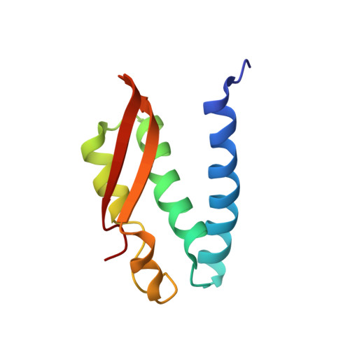
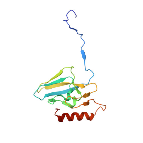
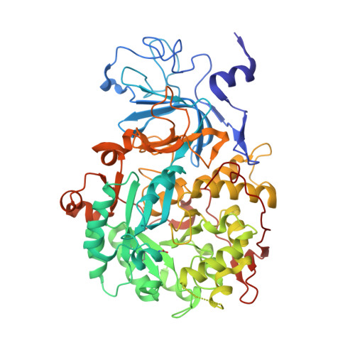
High-resolution cryo-EM structure of urease from the pathogen Yersinia enterocolitica.

Righetto, R.D.Anton, L.Adaixo, R.Jakob, R.P.Zivanov, J.Mahi, M.A.Ringler, P.Schwede, T.Maier, T.Stahlberg, H.

(2020) Nat Commun 11: 5101-5101

  • DOI: https://doi.org/10.1038/s41467-020-18870-2
  • Primary Citation of Related Structures:  
    6YL3

  • PubMed Abstract: 

    Urease converts urea into ammonia and carbon dioxide and makes urea available as a nitrogen source for all forms of life except animals. In human bacterial pathogens, ureases also aid in the invasion of acidic environments such as the stomach by raising the surrounding pH. Here, we report the structure of urease from the pathogen Yersinia enterocolitica at 2 Å resolution from cryo-electron microscopy. Y. enterocolitica urease is a dodecameric assembly of a trimer of three protein chains, ureA, ureB and ureC. The high data quality enables detailed visualization of the urease bimetal active site and of the impact of radiation damage. The obtained structure is of sufficient quality to support drug development efforts.


  • Organizational Affiliation

    Center for Cellular Imaging and NanoAnalytics, Biozentrum, University of Basel, Mattenstrasse 26, CH-4058, Basel, Switzerland.


Macromolecules
Find similar proteins by:  (by identity cutoff)  |  3D Structure
Entity ID: 1
MoleculeChains Sequence LengthOrganismDetailsImage
Urease subunit gamma100Yersinia enterocolitica W22703Mutation(s): 0 
EC: 3.5.1.5
UniProt
Find proteins for F4MWM9 (Yersinia enterocolitica W22703)
Explore F4MWM9 
Go to UniProtKB:  F4MWM9
Entity Groups  
Sequence Clusters30% Identity50% Identity70% Identity90% Identity95% Identity100% Identity
UniProt GroupF4MWM9
Sequence Annotations
Expand
  • Reference Sequence
Find similar proteins by:  (by identity cutoff)  |  3D Structure
Entity ID: 2
MoleculeChains Sequence LengthOrganismDetailsImage
Urease subunit beta132Yersinia enterocolitica W22703Mutation(s): 0 
EC: 3.5.1.5
UniProt
Find proteins for F4MWM8 (Yersinia enterocolitica W22703)
Explore F4MWM8 
Go to UniProtKB:  F4MWM8
Entity Groups  
Sequence Clusters30% Identity50% Identity70% Identity90% Identity95% Identity100% Identity
UniProt GroupF4MWM8
Sequence Annotations
Expand
  • Reference Sequence
Find similar proteins by:  (by identity cutoff)  |  3D Structure
Entity ID: 3
MoleculeChains Sequence LengthOrganismDetailsImage
Urease subunit alpha571Yersinia enterocolitica W22703Mutation(s): 0 
EC: 3.5.1.5
UniProt
Find proteins for F4MWM7 (Yersinia enterocolitica W22703)
Explore F4MWM7 
Go to UniProtKB:  F4MWM7
Entity Groups  
Sequence Clusters30% Identity50% Identity70% Identity90% Identity95% Identity100% Identity
UniProt GroupF4MWM7
Sequence Annotations
Expand
  • Reference Sequence
Small Molecules
Ligands 1 Unique
IDChains Name / Formula / InChI Key2D Diagram3D Interactions
NI
Query on NI

Download Ideal Coordinates CCD File 
AB [auth 1]
BB [auth 1]
CB [auth 4]
DB [auth 4]
EB [auth 7]
AB [auth 1],
BB [auth 1],
CB [auth 4],
DB [auth 4],
EB [auth 7],
FB [auth 7],
GB [auth U],
HB [auth U],
KA [auth C],
LA [auth C],
MA [auth F],
NA [auth F],
OA [auth I],
PA [auth I],
QA [auth L],
RA [auth L],
SA [auth O],
TA [auth O],
UA [auth R],
VA [auth R],
WA [auth V],
XA [auth V],
YA [auth Y],
ZA [auth Y]
NICKEL (II) ION
Ni
VEQPNABPJHWNSG-UHFFFAOYSA-N
Modified Residues  1 Unique
IDChains TypeFormula2D DiagramParent
KCX
Query on KCX
AA [auth 1]
C
DA [auth 4]
F
GA [auth 7]
AA [auth 1],
C,
DA [auth 4],
F,
GA [auth 7],
I,
JA [auth U],
L,
O,
R,
U [auth V],
X [auth Y]
L-PEPTIDE LINKINGC7 H14 N2 O4LYS
Experimental Data & Validation

Experimental Data

  • Method: ELECTRON MICROSCOPY
  • Resolution: 1.98 Å
  • Aggregation State: PARTICLE 
  • Reconstruction Method: SINGLE PARTICLE 
EM Software:
TaskSoftware PackageVersion
RECONSTRUCTIONRELION3.1
MODEL REFINEMENTPHENIX

Structure Validation

View Full Validation Report


Entry History & Funding Information

Deposition Data


Funding OrganizationLocationGrant Number
Swiss National Science FoundationSwitzerland--

Revision History  (Full details and data files)

  • Version 1.0: 2020-05-06
    Type: Initial release
  • Version 1.1: 2020-10-21
    Changes: Database references, Derived calculations