6YCZ

Plasmodium falciparum Myosin A delta-Nter, Post-Rigor state


Experimental Data Snapshot

  • Method: X-RAY DIFFRACTION
  • Resolution: 3.27 Å
  • R-Value Free: 0.227 
  • R-Value Work: 0.179 
  • R-Value Observed: 0.182 

wwPDB Validation   3D Report Full Report


Ligand Structure Quality Assessment 


This is version 1.1 of the entry. See complete history


Literature

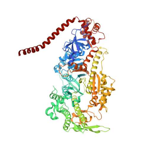
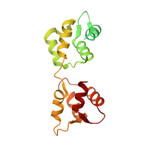
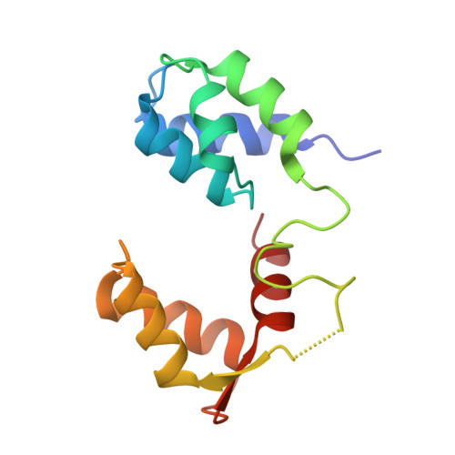
Full-length Plasmodium falciparum myosin A and essential light chain PfELC structures provide new anti-malarial targets.

Moussaoui, D.Robblee, J.P.Auguin, D.Krementsova, E.B.Haase, S.Blake, T.C.A.Baum, J.Robert-Paganin, J.Trybus, K.M.Houdusse, A.

(2020) Elife 9

  • DOI: https://doi.org/10.7554/eLife.60581
  • Primary Citation of Related Structures:  
    6YCX, 6YCY, 6YCZ

  • PubMed Abstract: 

    Parasites from the genus Plasmodium are the causative agents of malaria. The mobility, infectivity, and ultimately pathogenesis of Plasmodium falciparum rely on a macromolecular complex, called the glideosome. At the core of the glideosome is an essential and divergent Myosin A motor (PfMyoA), a first order drug target against malaria. Here, we present the full-length structure of PfMyoA in two states of its motor cycle. We report novel interactions that are essential for motor priming and the mode of recognition of its two light chains (PfELC and MTIP) by two degenerate IQ motifs. Kinetic and motility assays using PfMyoA variants, along with molecular dynamics, demonstrate how specific priming and atypical sequence adaptations tune the motor's mechano-chemical properties. Supported by evidence for an essential role of the PfELC in malaria pathogenesis, these structures provide a blueprint for the design of future anti-malarials targeting both the glideosome motor and its regulatory elements.


  • Organizational Affiliation

    Structural Motility, Institut Curie, Paris Université Sciences et Lettres, Sorbonne Université, CNRS UMR144, Paris, France.


Macromolecules
Find similar proteins by:  (by identity cutoff)  |  3D Structure
Entity ID: 1
MoleculeChains Sequence LengthOrganismDetailsImage
Myosin-A818Plasmodium falciparum 3D7Mutation(s): 0 
Gene Names: PF13_0233
UniProt
Find proteins for Q8IDR3 (Plasmodium falciparum (isolate 3D7))
Explore Q8IDR3 
Go to UniProtKB:  Q8IDR3
Entity Groups  
Sequence Clusters30% Identity50% Identity70% Identity90% Identity95% Identity100% Identity
UniProt GroupQ8IDR3
Sequence Annotations
Expand
  • Reference Sequence
Find similar proteins by:  (by identity cutoff)  |  3D Structure
Entity ID: 2
MoleculeChains Sequence LengthOrganismDetailsImage
Myosin A tail domain interacting protein204Plasmodium falciparum NF54Mutation(s): 0 
Gene Names: CK202_1873PFNF54_03975
UniProt
Find proteins for W7K1J7 (Plasmodium falciparum (isolate NF54))
Explore W7K1J7 
Go to UniProtKB:  W7K1J7
Entity Groups  
Sequence Clusters30% Identity50% Identity70% Identity90% Identity95% Identity100% Identity
UniProt GroupW7K1J7
Sequence Annotations
Expand
  • Reference Sequence
Find similar proteins by:  (by identity cutoff)  |  3D Structure
Entity ID: 3
MoleculeChains Sequence LengthOrganismDetailsImage
Uncharacterized protein134Plasmodium falciparum NF54Mutation(s): 0 
Gene Names: CK202_4702
UniProt
Find proteins for A0A2I0BQX1 (Plasmodium falciparum (isolate NF54))
Explore A0A2I0BQX1 
Go to UniProtKB:  A0A2I0BQX1
Entity Groups  
Sequence Clusters30% Identity50% Identity70% Identity90% Identity95% Identity100% Identity
UniProt GroupA0A2I0BQX1
Sequence Annotations
Expand
  • Reference Sequence
Small Molecules
Ligands 4 Unique
IDChains Name / Formula / InChI Key2D Diagram3D Interactions
ADP (Subject of Investigation/LOI)
Query on ADP

Download Ideal Coordinates CCD File 
H [auth A]ADENOSINE-5'-DIPHOSPHATE
C10 H15 N5 O10 P2
XTWYTFMLZFPYCI-KQYNXXCUSA-N
SO4
Query on SO4

Download Ideal Coordinates CCD File 
D [auth A],
E [auth A],
I [auth B]
SULFATE ION
O4 S
QAOWNCQODCNURD-UHFFFAOYSA-L
EDO
Query on EDO

Download Ideal Coordinates CCD File 
F [auth A],
J [auth C]
1,2-ETHANEDIOL
C2 H6 O2
LYCAIKOWRPUZTN-UHFFFAOYSA-N
MG (Subject of Investigation/LOI)
Query on MG

Download Ideal Coordinates CCD File 
G [auth A]MAGNESIUM ION
Mg
JLVVSXFLKOJNIY-UHFFFAOYSA-N
Experimental Data & Validation

Experimental Data

  • Method: X-RAY DIFFRACTION
  • Resolution: 3.27 Å
  • R-Value Free: 0.227 
  • R-Value Work: 0.179 
  • R-Value Observed: 0.182 
  • Space Group: P 21 21 21
Unit Cell:
Length ( Å )Angle ( ˚ )
a = 90.08α = 90
b = 114.43β = 90
c = 170.71γ = 90
Software Package:
Software NamePurpose
BUSTERrefinement
PDB_EXTRACTdata extraction
XDSdata reduction
XDSdata scaling
PHASERphasing

Structure Validation

View Full Validation Report


Ligand Structure Quality Assessment 


Entry History & Funding Information

Deposition Data


Funding OrganizationLocationGrant Number
National Institutes of Health/National Institute of General Medical Sciences (NIH/NIGMS)AI132378

Revision History  (Full details and data files)

  • Version 1.0: 2020-11-11
    Type: Initial release
  • Version 1.1: 2024-01-24
    Changes: Data collection, Database references, Refinement description