6Y8P

Crystal structure of SNAP-tag labeled with a benzyl-tetramethylrhodamine fluorophore


Experimental Data Snapshot

  • Method: X-RAY DIFFRACTION
  • Resolution: 2.30 Å
  • R-Value Free: 0.277 
  • R-Value Work: 0.236 
  • R-Value Observed: 0.237 

wwPDB Validation   3D Report Full Report


This is version 1.3 of the entry. See complete history


Literature

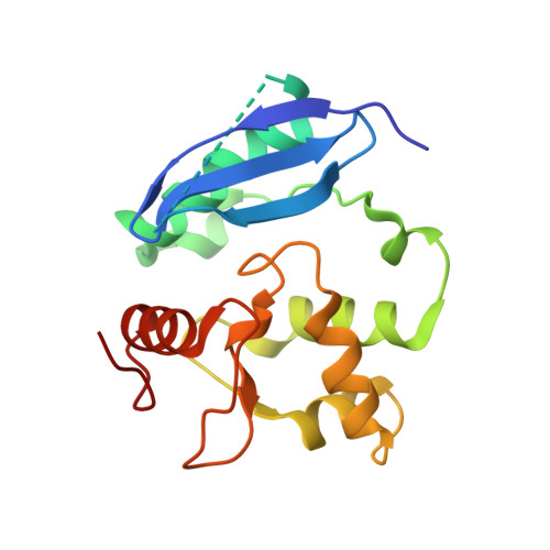
Kinetic and Structural Characterization of the Self-Labeling Protein Tags HaloTag7, SNAP-tag, and CLIP-tag.

Wilhelm, J.Kuhn, S.Tarnawski, M.Gotthard, G.Tunnermann, J.Tanzer, T.Karpenko, J.Mertes, N.Xue, L.Uhrig, U.Reinstein, J.Hiblot, J.Johnsson, K.

(2021) Biochemistry 60: 2560-2575

  • DOI: https://doi.org/10.1021/acs.biochem.1c00258
  • Primary Citation of Related Structures:  
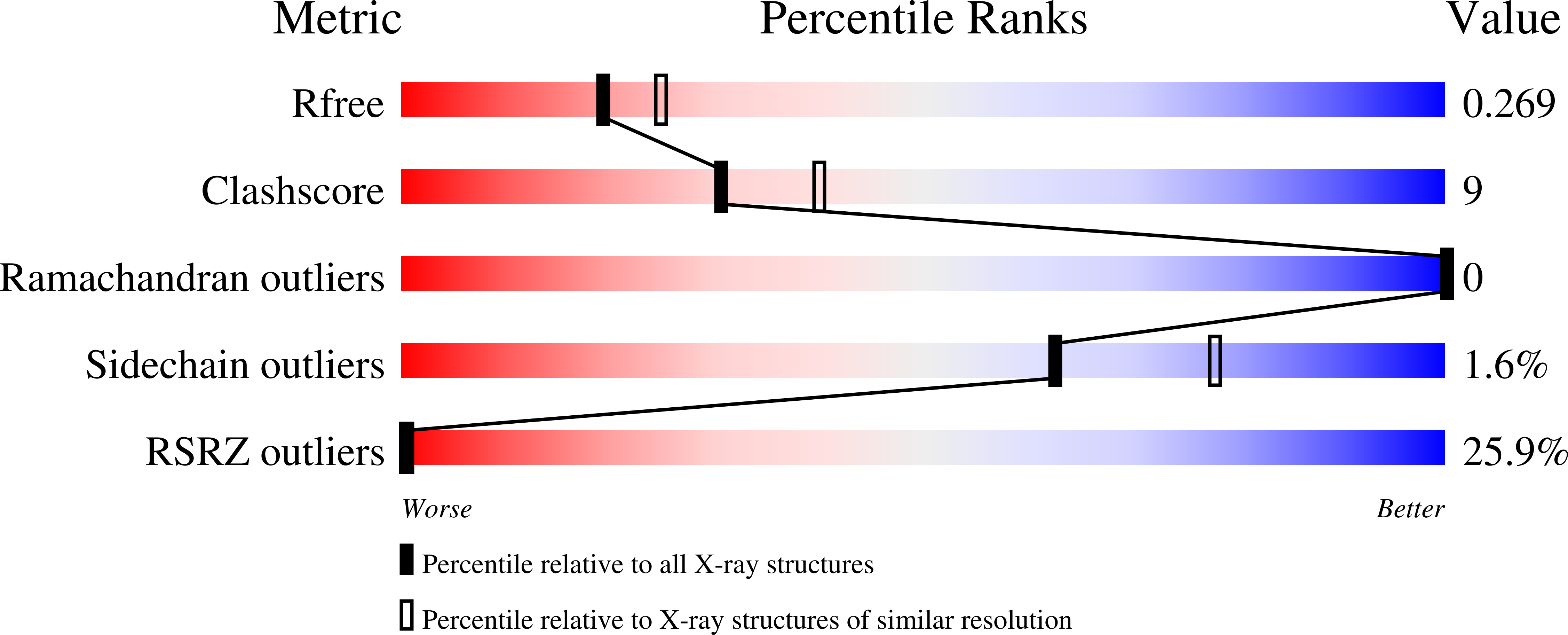
    6Y7A, 6Y7B, 6Y8P, 6ZCC

  • PubMed Abstract: 

    The self-labeling protein tags (SLPs) HaloTag7, SNAP-tag, and CLIP-tag allow the covalent labeling of fusion proteins with synthetic molecules for applications in bioimaging and biotechnology. To guide the selection of an SLP-substrate pair and provide guidelines for the design of substrates, we report a systematic and comparative study of the labeling kinetics and substrate specificities of HaloTag7, SNAP-tag, and CLIP-tag. HaloTag7 reaches almost diffusion-limited labeling rate constants with certain rhodamine substrates, which are more than 2 orders of magnitude higher than those of SNAP-tag for the corresponding substrates. SNAP-tag labeling rate constants, however, are less affected by the structure of the label than those of HaloTag7, which vary over 6 orders of magnitude for commonly employed substrates. Determining the crystal structures of HaloTag7 and SNAP-tag labeled with fluorescent substrates allowed us to rationalize their substrate preferences. We also demonstrate how these insights can be exploited to design substrates with improved labeling kinetics.


  • Organizational Affiliation

    Department of Chemical Biology, Max Planck Institute for Medical Research, 69120 Heidelberg, Germany.


Macromolecules
Find similar proteins by:  (by identity cutoff)  |  3D Structure
Entity ID: 1
MoleculeChains Sequence LengthOrganismDetailsImage
O6-alkylguanine-DNA alkyltransferase mutant177Homo sapiensMutation(s): 0 
UniProt
Find proteins for E5BBQ0 (Homo sapiens)
Explore E5BBQ0 
Go to UniProtKB:  E5BBQ0
Entity Groups  
Sequence Clusters30% Identity50% Identity70% Identity90% Identity95% Identity100% Identity
UniProt GroupE5BBQ0
Sequence Annotations
Expand
  • Reference Sequence
Small Molecules
Ligands 3 Unique
IDChains Name / Formula / InChI Key2D Diagram3D Interactions
OGQ (Subject of Investigation/LOI)
Query on OGQ

Download Ideal Coordinates CCD File 
C [auth A][9-[2-carboxy-5-[(4-methylphenyl)methylcarbamoyl]phenyl]-6-(dimethylamino)xanthen-3-ylidene]-dimethyl-azanium
C33 H32 N3 O4
VRTFWVCUZQBQCQ-UHFFFAOYSA-O
ZN
Query on ZN

Download Ideal Coordinates CCD File 
B [auth A]ZINC ION
Zn
PTFCDOFLOPIGGS-UHFFFAOYSA-N
EDO
Query on EDO

Download Ideal Coordinates CCD File 
D [auth A]1,2-ETHANEDIOL
C2 H6 O2
LYCAIKOWRPUZTN-UHFFFAOYSA-N
Experimental Data & Validation

Experimental Data

  • Method: X-RAY DIFFRACTION
  • Resolution: 2.30 Å
  • R-Value Free: 0.277 
  • R-Value Work: 0.236 
  • R-Value Observed: 0.237 
  • Space Group: P 32 2 1
Unit Cell:
Length ( Å )Angle ( ˚ )
a = 65.515α = 90
b = 65.515β = 90
c = 97.067γ = 120
Software Package:
Software NamePurpose
PHENIXrefinement
PDB_EXTRACTdata extraction
XDSdata reduction
XSCALEdata scaling
PHASERphasing

Structure Validation

View Full Validation Report


Entry History 

Deposition Data

Revision History  (Full details and data files)

  • Version 1.0: 2021-03-31
    Type: Initial release
  • Version 1.1: 2021-10-27
    Changes: Data collection, Database references
  • Version 1.2: 2021-11-10
    Changes: Data collection, Database references
  • Version 1.3: 2024-01-24
    Changes: Data collection, Database references, Refinement description