6Y68

Structure of Maporal virus envelope glycoprotein Gc in postfusion conformation


Experimental Data Snapshot

  • Method: X-RAY DIFFRACTION
  • Resolution: 2.40 Å
  • R-Value Free: 0.236 
  • R-Value Work: 0.196 
  • R-Value Observed: 0.198 

wwPDB Validation   3D Report Full Report


This is version 1.2 of the entry. See complete history


Literature

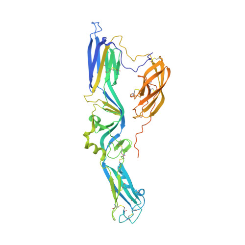
The Hantavirus Surface Glycoprotein Lattice and Its Fusion Control Mechanism.

Serris, A.Stass, R.Bignon, E.A.Muena, N.A.Manuguerra, J.C.Jangra, R.K.Li, S.Chandran, K.Tischler, N.D.Huiskonen, J.T.Rey, F.A.Guardado-Calvo, P.

(2020) Cell 183: 442

  • DOI: https://doi.org/10.1016/j.cell.2020.08.023
  • Primary Citation of Related Structures:  
    6Y5F, 6Y5W, 6Y62, 6Y68, 6Y6P, 6Y6Q, 6YRB, 6YRQ, 6ZJM

  • PubMed Abstract: 

    Hantaviruses are rodent-borne viruses causing serious zoonotic outbreaks worldwide for which no treatment is available. Hantavirus particles are pleomorphic and display a characteristic square surface lattice. The envelope glycoproteins Gn and Gc form heterodimers that further assemble into tetrameric spikes, the lattice building blocks. The glycoproteins, which are the sole targets of neutralizing antibodies, drive virus entry via receptor-mediated endocytosis and endosomal membrane fusion. Here we describe the high-resolution X-ray structures of the heterodimer of Gc and the Gn head and of the homotetrameric Gn base. Docking them into an 11.4-Å-resolution cryoelectron tomography map of the hantavirus surface accounted for the complete extramembrane portion of the viral glycoprotein shell and allowed a detailed description of the surface organization of these pleomorphic virions. Our results, which further revealed a built-in mechanism controlling Gc membrane insertion for fusion, pave the way for immunogen design to protect against pathogenic hantaviruses.


  • Organizational Affiliation

    Institut Pasteur, Structural Virology Unit, and CNRS UMR 3569, Paris, France.


Macromolecules
Find similar proteins by:  (by identity cutoff)  |  3D Structure
Entity ID: 1
MoleculeChains Sequence LengthOrganismDetailsImage
Envelope polyprotein494Maporal virusMutation(s): 0 
UniProt
Find proteins for Q5MYC0 (Maporal virus)
Explore Q5MYC0 
Go to UniProtKB:  Q5MYC0
Entity Groups  
Sequence Clusters30% Identity50% Identity70% Identity90% Identity95% Identity100% Identity
UniProt GroupQ5MYC0
Sequence Annotations
Expand
  • Reference Sequence
Oligosaccharides

Help

Entity ID: 2
MoleculeChains Length2D Diagram Glycosylation3D Interactions
alpha-D-mannopyranose-(1-3)-beta-D-mannopyranose-(1-4)-2-acetamido-2-deoxy-beta-D-glucopyranose-(1-4)-2-acetamido-2-deoxy-beta-D-glucopyranose
B
4N-Glycosylation
Glycosylation Resources
GlyTouCan:  G81315DD
GlyCosmos:  G81315DD
GlyGen:  G81315DD
Experimental Data & Validation

Experimental Data

  • Method: X-RAY DIFFRACTION
  • Resolution: 2.40 Å
  • R-Value Free: 0.236 
  • R-Value Work: 0.196 
  • R-Value Observed: 0.198 
  • Space Group: H 3 2
Unit Cell:
Length ( Å )Angle ( ˚ )
a = 90.348α = 90
b = 90.348β = 90
c = 506.38γ = 120
Software Package:
Software NamePurpose
XDSdata reduction
Aimlessdata scaling
PHASERphasing
PHENIXrefinement
PDB_EXTRACTdata extraction

Structure Validation

View Full Validation Report


Entry History & Funding Information

Deposition Data


Funding OrganizationLocationGrant Number
French National Research AgencyFranceANR-18-CE11-0011

Revision History  (Full details and data files)

  • Version 1.0: 2020-10-14
    Type: Initial release
  • Version 1.1: 2020-10-28
    Changes: Database references
  • Version 1.2: 2024-01-24
    Changes: Data collection, Database references, Refinement description