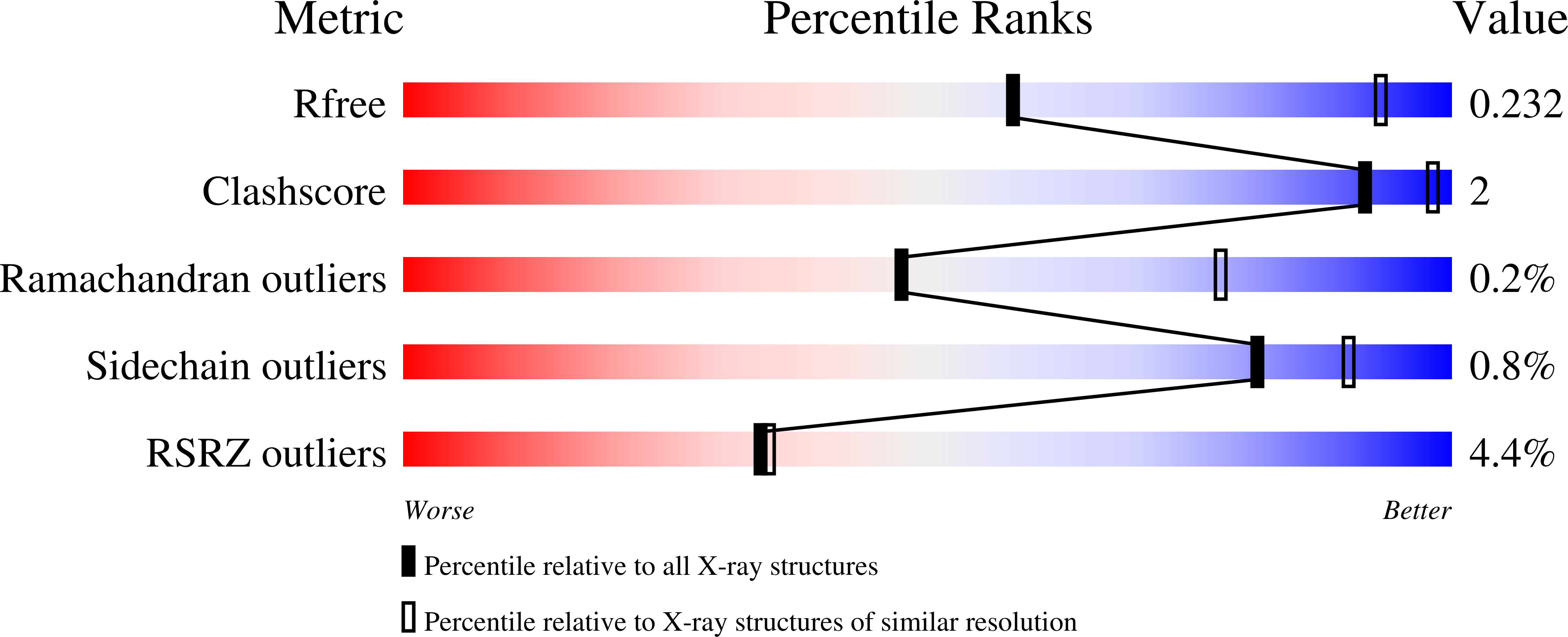
6Y4M

Structure of Tubulin Tyrosine Ligase in Complex with Tb111


Experimental Data Snapshot

  • Method: X-RAY DIFFRACTION
  • Resolution: 3.34 Å
  • R-Value Free: 0.234 
  • R-Value Work: 0.207 
  • R-Value Observed: 0.208 

wwPDB Validation   3D Report Full Report


Ligand Structure Quality Assessment 


This is version 1.1 of the entry. See complete history


Literature

Design, Synthesis, and Biological Evaluation of Tubulysin Analogues, Linker-Drugs, and Antibody-Drug Conjugates, Insights into Structure-Activity Relationships, and Tubulysin-Tubulin Binding Derived from X-ray Crystallographic Analysis.

Nicolaou, K.C.Pan, S.Pulukuri, K.K.Ye, Q.Rigol, S.Erande, R.D.Vourloumis, D.Nocek, B.P.Munneke, S.Lyssikatos, J.Valdiosera, A.Gu, C.Lin, B.Sarvaiaya, H.Trinidad, J.Sandoval, J.Lee, C.Hammond, M.Aujay, M.Taylor, N.Pysz, M.Purcell, J.W.Gavrilyuk, J.

(2021) J Org Chem 86: 3377-3421

  • DOI: https://doi.org/10.1021/acs.joc.0c02755
  • Primary Citation of Related Structures:  
    6Y4M, 6Y4N

  • PubMed Abstract: 

    Molecular design, synthesis, and biological evaluation of tubulysin analogues, linker-drugs, and antibody-drug conjugates are described. Among the new discoveries reported is the identification of new potent analogues within the tubulysin family that carry a C11 alkyl ether substituent, rather than the usual ester structural motif at that position, a fact that endows the former with higher plasma stability than that of the latter. Also described herein are X-ray crystallographic analysis studies of two tubulin-tubulysin complexes formed within the α/β interface between two tubulin heterodimers and two highly potent tubulysin analogues, one of which exhibited a different binding mode to the one previously reported for tubulysin M. The X-ray crystallographic analysis-derived new insights into the binding modes of these tubulysin analogues explain their potencies and provide inspiration for further design, synthesis, and biological investigations within this class of antitumor agents. A number of these analogues were conjugated as payloads with appropriate linkers at different sites allowing their attachment onto targeting antibodies for cancer therapies. A number of such antibody-drug conjugates were constructed and tested, both in vivo and in vitro, leading to the identification of at least one promising ADC (Herceptin- LD3 ), warranting further investigations.


  • Organizational Affiliation

    Department of Chemistry, BioScience Research Collaborative, Rice University, 6100 Main Street, Houston, Texas 77005, United States.


Macromolecules
Find similar proteins by:  (by identity cutoff)  |  3D Structure
Entity ID: 1
MoleculeChains Sequence LengthOrganismDetailsImage
Tubulin alpha-1B chain
A, C
451Sus scrofaMutation(s): 0 
UniProt
Find proteins for Q2XVP4 (Sus scrofa)
Explore Q2XVP4 
Go to UniProtKB:  Q2XVP4
Entity Groups  
Sequence Clusters30% Identity50% Identity70% Identity90% Identity95% Identity100% Identity
UniProt GroupQ2XVP4
Sequence Annotations
Expand
  • Reference Sequence
Find similar proteins by:  (by identity cutoff)  |  3D Structure
Entity ID: 2
MoleculeChains Sequence LengthOrganismDetailsImage
Tubulin beta chain
B, D
445Sus scrofaMutation(s): 0 
UniProt
Find proteins for P02554 (Sus scrofa)
Explore P02554 
Go to UniProtKB:  P02554
Entity Groups  
Sequence Clusters30% Identity50% Identity70% Identity90% Identity95% Identity100% Identity
UniProt GroupP02554
Sequence Annotations
Expand
  • Reference Sequence
Find similar proteins by:  (by identity cutoff)  |  3D Structure
Entity ID: 3
MoleculeChains Sequence LengthOrganismDetailsImage
Stathmin-4143Rattus norvegicusMutation(s): 0 
Gene Names: Stmn4
UniProt
Find proteins for P63043 (Rattus norvegicus)
Explore P63043 
Go to UniProtKB:  P63043
Entity Groups  
Sequence Clusters30% Identity50% Identity70% Identity90% Identity95% Identity100% Identity
UniProt GroupP63043
Sequence Annotations
Expand
  • Reference Sequence
Find similar proteins by:  (by identity cutoff)  |  3D Structure
Entity ID: 4
MoleculeChains Sequence LengthOrganismDetailsImage
Tubulin-Tyrosine Ligase384Gallus gallusMutation(s): 0 
Gene Names: TTL
Entity Groups  
Sequence Clusters30% Identity50% Identity70% Identity90% Identity95% Identity100% Identity
Sequence Annotations
Expand
  • Reference Sequence
Small Molecules
Ligands 11 Unique
IDChains Name / Formula / InChI Key2D Diagram3D Interactions
GTP
Query on GTP

Download Ideal Coordinates CCD File 
G [auth A],
P [auth C],
U [auth D]
GUANOSINE-5'-TRIPHOSPHATE
C10 H16 N5 O14 P3
XKMLYUALXHKNFT-UUOKFMHZSA-N
ACP
Query on ACP

Download Ideal Coordinates CCD File 
W [auth F]PHOSPHOMETHYLPHOSPHONIC ACID ADENYLATE ESTER
C11 H18 N5 O12 P3
UFZTZBNSLXELAL-IOSLPCCCSA-N
GDP
Query on GDP

Download Ideal Coordinates CCD File 
J [auth B]GUANOSINE-5'-DIPHOSPHATE
C10 H15 N5 O11 P2
QGWNDRXFNXRZMB-UUOKFMHZSA-N
O9H
Query on O9H

Download Ideal Coordinates CCD File 
N [auth B][(1~{R},3~{R})-1-acetyloxy-1-[4-methanoyl-5-(2-phenylmethoxyethyl)-2,3-dihydro-1,3-thiazol-2-yl]-4-methyl-pentan-3-yl]-methyl-azanium
C22 H33 N2 O4 S
PGOBLOKJRKQHRJ-UZKOGDIHSA-O
OH5
Query on OH5

Download Ideal Coordinates CCD File 
O [auth B](2~{S},4~{R})-4-azanyl-2-methyl-5-phenyl-pentanoic acid
C12 H17 N O2
TVHNWAKCVXFPNB-GXSJLCMTSA-N
MES
Query on MES

Download Ideal Coordinates CCD File 
L [auth B]2-(N-MORPHOLINO)-ETHANESULFONIC ACID
C6 H13 N O4 S
SXGZJKUKBWWHRA-UHFFFAOYSA-N
PGE
Query on PGE

Download Ideal Coordinates CCD File 
S [auth C]TRIETHYLENE GLYCOL
C6 H14 O4
ZIBGPFATKBEMQZ-UHFFFAOYSA-N
O9B
Query on O9B

Download Ideal Coordinates CCD File 
M [auth B](2~{R})-1-methylpiperidine-2-carboxylic acid
C7 H13 N O2
BPSLZWSRHTULGU-ZCFIWIBFSA-N
VAL
Query on VAL

Download Ideal Coordinates CCD File 
T [auth C]VALINE
C5 H11 N O2
KZSNJWFQEVHDMF-BYPYZUCNSA-N
CA
Query on CA

Download Ideal Coordinates CCD File 
I [auth A],
R [auth C]
CALCIUM ION
Ca
BHPQYMZQTOCNFJ-UHFFFAOYSA-N
MG
Query on MG

Download Ideal Coordinates CCD File 
H [auth A],
K [auth B],
Q [auth C],
V [auth D]
MAGNESIUM ION
Mg
JLVVSXFLKOJNIY-UHFFFAOYSA-N
Experimental Data & Validation

Experimental Data

  • Method: X-RAY DIFFRACTION
  • Resolution: 3.34 Å
  • R-Value Free: 0.234 
  • R-Value Work: 0.207 
  • R-Value Observed: 0.208 
  • Space Group: P 21 21 21
Unit Cell:
Length ( Å )Angle ( ˚ )
a = 103.961α = 90
b = 152.59β = 90
c = 185.989γ = 90
Software Package:
Software NamePurpose
MOSFLMdata reduction
SCALAdata scaling
REFMACrefinement
PDB_EXTRACTdata extraction

Structure Validation

View Full Validation Report


Ligand Structure Quality Assessment 


Entry History 

Deposition Data

Revision History  (Full details and data files)

  • Version 1.0: 2021-03-31
    Type: Initial release
  • Version 1.1: 2022-11-23
    Changes: Database references