6Y3G

Crystal structure of phenylalanine tRNA from Escherichia coli


Experimental Data Snapshot

  • Method: X-RAY DIFFRACTION
  • Resolution: 3.10 Å
  • R-Value Free: 0.236 
  • R-Value Work: 0.215 
  • R-Value Observed: 0.216 

wwPDB Validation   3D Report Full Report


This is version 1.1 of the entry. See complete history


Literature

Structural basis of the interaction between cyclodipeptide synthases and aminoacylated tRNA substrates.

Bourgeois, G.Seguin, J.Babin, M.Gondry, M.Mechulam, Y.Schmitt, E.

(2020) RNA 26: 1589-1602

  • DOI: https://doi.org/10.1261/rna.075184.120
  • Primary Citation of Related Structures:  
    6Y3G, 6Y4B

  • PubMed Abstract: 

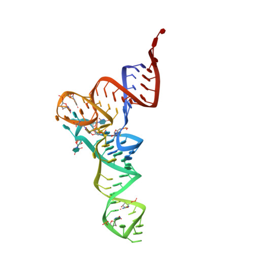
    Cyclodipeptide synthases (CDPSs) catalyze the synthesis of various cyclodipeptides by using two aminoacyl-tRNA (aa-tRNA) substrates in a sequential mechanism. Here, we studied binding of phenylalanyl-tRNA Phe to the CDPS from Candidatus Glomeribacter gigasporarum ( Cglo -CDPS) by gel filtration and electrophoretic mobility shift assay. We determined the crystal structure of the Cglo -CDPS:Phe-tRNA Phe complex to 5 Å resolution and further studied it in solution using small-angle X-ray scattering (SAXS). The data show that the major groove of the acceptor stem of the aa-tRNA interacts with the enzyme through the basic β2 and β7 strands of CDPSs belonging to the XYP subfamily. A bending of the CCA extremity enables the amino acid moiety to be positioned in the P1 pocket while the terminal A76 adenosine occupies the P2 pocket. Such a positioning indicates that the present structure illustrates the binding of the first aa-tRNA. In cells, CDPSs and the elongation factor EF-Tu share aminoacylated tRNAs as substrates. The present study shows that CDPSs and EF-Tu interact with opposite sides of tRNA. This may explain how CDPSs hijack aa-tRNAs from canonical ribosomal protein synthesis.


  • Organizational Affiliation

    Laboratoire de Biologie Structurale de la Cellule, BIOC, Ecole polytechnique, CNRS, Institut Polytechnique de Paris, 91128 Palaiseau cedex, France.


Macromolecules
Find similar nucleic acids by:  (by identity cutoff)  |  3D Structure
Entity ID: 1
MoleculeChains LengthOrganismImage
RNA (75-MER)A [auth F]76Escherichia coli
Sequence Annotations
Expand
  • Reference Sequence
Small Molecules
Ligands 3 Unique
IDChains Name / Formula / InChI Key2D Diagram3D Interactions
GOL
Query on GOL

Download Ideal Coordinates CCD File 
M [auth F]GLYCEROL
C3 H8 O3
PEDCQBHIVMGVHV-UHFFFAOYSA-N
GAI
Query on GAI

Download Ideal Coordinates CCD File 
B [auth F],
C [auth F],
D [auth F],
E [auth F]
GUANIDINE
C H5 N3
ZRALSGWEFCBTJO-UHFFFAOYSA-N
CA
Query on CA

Download Ideal Coordinates CCD File 
F
G [auth F]
H [auth F]
I [auth F]
J [auth F]
F,
G [auth F],
H [auth F],
I [auth F],
J [auth F],
K [auth F],
L [auth F]
CALCIUM ION
Ca
BHPQYMZQTOCNFJ-UHFFFAOYSA-N
Experimental Data & Validation

Experimental Data

  • Method: X-RAY DIFFRACTION
  • Resolution: 3.10 Å
  • R-Value Free: 0.236 
  • R-Value Work: 0.215 
  • R-Value Observed: 0.216 
  • Space Group: P 64 2 2
Unit Cell:
Length ( Å )Angle ( ˚ )
a = 109.618α = 90
b = 109.618β = 90
c = 138.518γ = 120
Software Package:
Software NamePurpose
XDSdata reduction
PHENIXrefinement
XDSdata reduction
XDSdata scaling
PHASERphasing

Structure Validation

View Full Validation Report


Entry History & Funding Information

Deposition Data


Funding OrganizationLocationGrant Number
French National Research AgencyFrance14CE090021

Revision History  (Full details and data files)

  • Version 1.0: 2020-12-30
    Type: Initial release
  • Version 1.1: 2024-01-24
    Changes: Data collection, Database references, Refinement description, Structure summary