6Y38

Crystal structure of Whirlin PDZ3 in complex with Myosin 15a C-terminal PDZ binding motif peptide


Experimental Data Snapshot

  • Method: X-RAY DIFFRACTION
  • Resolution: 1.70 Å
  • R-Value Free: 0.231 
  • R-Value Work: 0.207 

wwPDB Validation   3D Report Full Report


This is version 1.2 of the entry. See complete history


Literature

Deciphering the Unexpected Binding Capacity of the Third PDZ Domain of Whirlin to Various Cochlear Hair Cell Partners.

Zhu, Y.Delhommel, F.Cordier, F.Luchow, S.Mechaly, A.Colcombet-Cazenave, B.Girault, V.Pepermans, E.Bahloul, A.Gautier, C.Brule, S.Raynal, B.Hoos, S.Haouz, A.Caillet-Saguy, C.Ivarsson, Y.Wolff, N.

(2020) J Mol Biol 432: 5920-5937

  • DOI: https://doi.org/10.1016/j.jmb.2020.09.012
  • Primary Citation of Related Structures:  
    6Y38, 6Y9N, 6Y9O, 6Y9P, 6Y9Q

  • PubMed Abstract: 

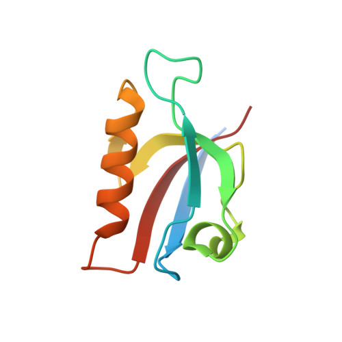
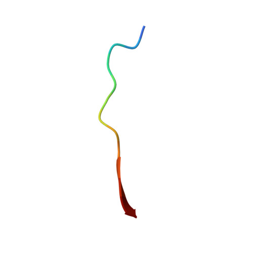
    Hearing is a mechanical and neurochemical process, which occurs in the hair cells of inner ear that converts the sound vibrations into electrical signals transmitted to the brain. The multi-PDZ scaffolding protein whirlin plays a critical role in the formation and function of stereocilia exposed at the surface of hair cells. In this article, we reported seven stereociliary proteins that encode PDZ binding motifs (PBM) and interact with whirlin PDZ3, where four of them are first reported. We solved the atomic resolution structures of complexes between whirlin PDZ3 and the PBMs of myosin 15a, CASK, harmonin a1 and taperin. Interestingly, the PBM of CASK and taperin are rare non-canonical PBM, which are not localized at the extreme C terminus. This large capacity to accommodate various partners could be related to the distinct functions of whirlin at different stages of the hair cell development.


  • Organizational Affiliation

    Unité Récepteurs-Canaux, Institut Pasteur, 75015 Paris, France; Complexité du Vivant, Sorbonne Université, 75005 Paris, France.


Macromolecules
Find similar proteins by:  (by identity cutoff)  |  3D Structure
Entity ID: 1
MoleculeChains Sequence LengthOrganismDetailsImage
Whirlin
A, B
102Mus musculusMutation(s): 0 
Gene Names: WhrnDfnb31Kiaa1526
UniProt & NIH Common Fund Data Resources
Find proteins for Q80VW5 (Mus musculus)
Explore Q80VW5 
Go to UniProtKB:  Q80VW5
IMPC:  MGI:2682003
Entity Groups  
Sequence Clusters30% Identity50% Identity70% Identity90% Identity95% Identity100% Identity
UniProt GroupQ80VW5
Sequence Annotations
Expand
  • Reference Sequence

Find similar proteins by:  Sequence   |   3D Structure  

Entity ID: 2
MoleculeChains Sequence LengthOrganismDetailsImage
Chains: C,D
C, D
13Mus musculusMutation(s): 0 
UniProt & NIH Common Fund Data Resources
Find proteins for Q9QZZ4 (Mus musculus)
Explore Q9QZZ4 
Go to UniProtKB:  Q9QZZ4
IMPC:  MGI:1261811
Entity Groups  
Sequence Clusters30% Identity50% Identity70% Identity90% Identity95% Identity100% Identity
UniProt GroupQ9QZZ4
Sequence Annotations
Expand
  • Reference Sequence
Experimental Data & Validation

Experimental Data

  • Method: X-RAY DIFFRACTION
  • Resolution: 1.70 Å
  • R-Value Free: 0.231 
  • R-Value Work: 0.207 
  • Space Group: P 21 21 2
Unit Cell:
Length ( Å )Angle ( ˚ )
a = 54.14α = 90
b = 112.92β = 90
c = 34.63γ = 90
Software Package:
Software NamePurpose
BUSTERrefinement
XDSdata reduction
Aimlessdata scaling
PHASERphasing

Structure Validation

View Full Validation Report


Entry History & Funding Information

Deposition Data


Funding OrganizationLocationGrant Number
Marie Sklodowska-Curie Actions, FragNET ITNFrance675341

Revision History  (Full details and data files)

  • Version 1.0: 2020-10-07
    Type: Initial release
  • Version 1.1: 2020-11-11
    Changes: Database references
  • Version 1.2: 2024-01-24
    Changes: Data collection, Database references, Refinement description