Guanidinium binding to proteins: The intriguing effects on the D1 and D2 domains of Thermotoga maritima Arginine Binding Protein and a comprehensive analysis of the Protein Data Bank.
Cozzolino, S., Balasco, N., Vigorita, M., Ruggiero, A., Smaldone, G., Del Vecchio, P., Vitagliano, L., Graziano, G.(2020) Int J Biol Macromol 163: 375-385
- PubMed: 32629051
- DOI: https://doi.org/10.1016/j.ijbiomac.2020.06.290
- Primary Citation of Related Structures:
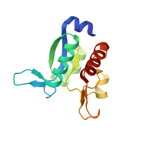
6Y16 - PubMed Abstract:
Thermotoga maritima Arginine Binding Protein has been extensively characterized because of its peculiar features and its possible use as a biosensor. In this characterization, deletion of the C-terminal helix to obtain the monomeric protein TmArgBP 20-233 and dissection of the monomer in its two domains, D1 and D2, have been performed. In the present study the stability of these three forms against guanidinium chloride is investigated by means of circular dichroism and differential scanning calorimetry measurements. All three proteins show a high conformational stability; moreover, D1 shows an unusual behavior in the presence of low concentrations of guanidinium chloride. This finding has led us to investigate a possible binding interaction by means of isothermal titration calorimetry and X-ray crystallography; the results indicate that D1 is able to bind the guanidinium ion (GuH + ), due to its similarity with the arginine terminal moiety. The analysis of the structural and dynamic properties of the D1-GuH + complex indicates that the protein binds the ligand through multiple and diversified interactions. An exhaustive survey of the binding modes of GuH + to proteins indicates that this is a rather common feature. These observations provide interesting insights into the effects that GuH + is able to induce in protein structures.
Organizational Affiliation:
Department of Chemical Sciences, University of Naples Federico II, via Cintia, 80126 Napoli, Italy.