6XWK

Crystal structure of Phormidium rubidum phycocyanin


Experimental Data Snapshot

  • Method: X-RAY DIFFRACTION
  • Resolution: 1.17 Å
  • R-Value Free: 0.145 
  • R-Value Work: 0.119 

wwPDB Validation   3D Report Full Report


Ligand Structure Quality Assessment 


This is version 1.2 of the entry. See complete history


Literature

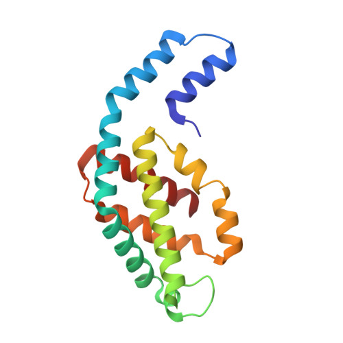
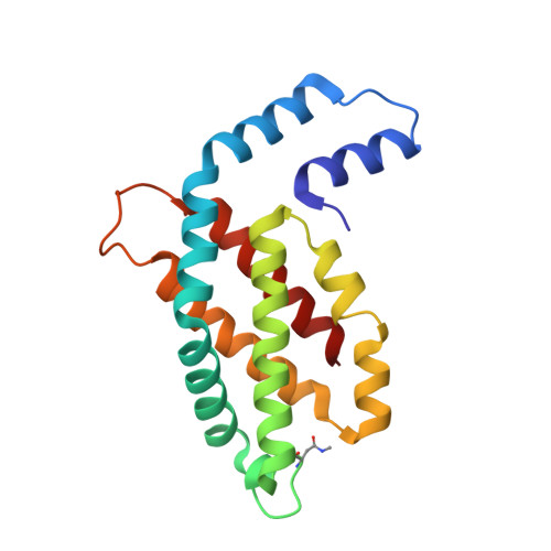
Revisiting high-resolution crystal structure of Phormidium rubidum phycocyanin.

Sonani, R.R.Roszak, A.W.Liu, H.Gross, M.L.Blankenship, R.E.Madamwar, D.Cogdell, R.J.

(2020) Photosynth Res 144: 349-360

  • DOI: https://doi.org/10.1007/s11120-020-00746-7
  • Primary Citation of Related Structures:  
    6XWK

  • PubMed Abstract: 

    The crystal structure of phycocyanin (pr-PC) isolated from Phormidium rubidum A09DM (P. rubidum) is described at a resolution of 1.17 Å. Electron density maps derived from crystallographic data showed many clear differences in amino acid sequences when compared with the previously obtained gene-derived sequences. The differences were found in 57 positions (30 in α-subunit and 27 in β-subunit of pr-PC), in which all residues except one (β145Arg) are not interacting with the three phycocyanobilin chromophores. Highly purified pr-PC was then sequenced by mass spectrometry (MS) using LC-MS/MS. The MS data were analyzed using two independent proteomic search engines. As a result of this analysis, complete agreement between the polypeptide sequences and the electron density maps was obtained. We attribute the difference to multiple genes in the bacterium encoding the phycocyanin apoproteins and that the gene sequencing sequenced the wrong ones. We are not implying that protein sequencing by mass spectrometry is more accurate than that of gene sequencing. The final 1.17 Å structure of pr-PC allows the chromophore interactions with the protein to be described with high accuracy.


  • Organizational Affiliation

    Post-Graduate Department of Biosciences, Sardar Patel University, Bakrol, Anand, Gujarat, 388 315, India.


Macromolecules
Find similar proteins by:  (by identity cutoff)  |  3D Structure
Entity ID: 1
MoleculeChains Sequence LengthOrganismDetailsImage
Phycocyanin alpha subunitA [auth AAA]162Phormidium rubidum A09DMMutation(s): 0 
Entity Groups  
Sequence Clusters30% Identity50% Identity70% Identity90% Identity95% Identity100% Identity
Sequence Annotations
Expand
  • Reference Sequence
Find similar proteins by:  (by identity cutoff)  |  3D Structure
Entity ID: 2
MoleculeChains Sequence LengthOrganismDetailsImage
Phycocyanin beta subunitB [auth BBB]172Phormidium rubidum A09DMMutation(s): 0 
Entity Groups  
Sequence Clusters30% Identity50% Identity70% Identity90% Identity95% Identity100% Identity
Sequence Annotations
Expand
  • Reference Sequence
Small Molecules
Ligands 5 Unique
IDChains Name / Formula / InChI Key2D Diagram3D Interactions
CYC (Subject of Investigation/LOI)
Query on CYC

Download Ideal Coordinates CCD File 
C [auth AAA],
M [auth BBB],
N [auth BBB]
PHYCOCYANOBILIN
C33 H40 N4 O6
VXTXPYZGDQPMHK-GMXXPEQVSA-N
PGE
Query on PGE

Download Ideal Coordinates CCD File 
O [auth BBB]TRIETHYLENE GLYCOL
C6 H14 O4
ZIBGPFATKBEMQZ-UHFFFAOYSA-N
PEG
Query on PEG

Download Ideal Coordinates CCD File 
D [auth AAA]DI(HYDROXYETHYL)ETHER
C4 H10 O3
MTHSVFCYNBDYFN-UHFFFAOYSA-N
IMD
Query on IMD

Download Ideal Coordinates CCD File 
J [auth AAA],
K [auth AAA],
P [auth BBB],
Q [auth BBB]
IMIDAZOLE
C3 H5 N2
RAXXELZNTBOGNW-UHFFFAOYSA-O
EDO
Query on EDO

Download Ideal Coordinates CCD File 
E [auth AAA]
F [auth AAA]
G [auth AAA]
H [auth AAA]
I [auth AAA]
E [auth AAA],
F [auth AAA],
G [auth AAA],
H [auth AAA],
I [auth AAA],
L [auth AAA]
1,2-ETHANEDIOL
C2 H6 O2
LYCAIKOWRPUZTN-UHFFFAOYSA-N
Modified Residues  1 Unique
IDChains TypeFormula2D DiagramParent
MEN
Query on MEN
B [auth BBB]L-PEPTIDE LINKINGC5 H10 N2 O3ASN
Experimental Data & Validation

Experimental Data

  • Method: X-RAY DIFFRACTION
  • Resolution: 1.17 Å
  • R-Value Free: 0.145 
  • R-Value Work: 0.119 
  • Space Group: P 63
Unit Cell:
Length ( Å )Angle ( ˚ )
a = 106.32α = 90
b = 106.32β = 90
c = 58.67γ = 120
Software Package:
Software NamePurpose
REFMACrefinement
XDSdata reduction
Aimlessdata scaling
PHASERphasing

Structure Validation

View Full Validation Report


Ligand Structure Quality Assessment 


Entry History & Funding Information

Deposition Data


Funding OrganizationLocationGrant Number
Department of Energy (DOE, United States)United StatesDE-SC0001035

Revision History  (Full details and data files)

  • Version 1.0: 2020-04-29
    Type: Initial release
  • Version 1.1: 2020-06-03
    Changes: Database references
  • Version 1.2: 2024-01-24
    Changes: Data collection, Database references, Derived calculations, Refinement description