6XVD

Crystal structure of complex of urokinase and a upain-1 variant(W3F) in pH7.4 condition


Experimental Data Snapshot

  • Method: X-RAY DIFFRACTION
  • Resolution: 1.40 Å
  • R-Value Free: 0.203 
  • R-Value Work: 0.186 
  • R-Value Observed: 0.187 

wwPDB Validation   3D Report Full Report


This is version 1.2 of the entry. See complete history


Literature

Insight to the residue in P2 position prevents the peptide inhibitor from being hydrolyzed by serine proteases.

Xue, G.Xie, X.Zhou, Y.Yuan, C.Huang, M.Jiang, L.

(2020) Biosci Biotechnol Biochem 84: 1153-1159

  • DOI: https://doi.org/10.1080/09168451.2020.1723405
  • Primary Citation of Related Structures:  
    6XVD

  • PubMed Abstract: 

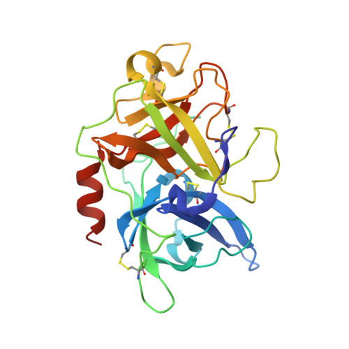
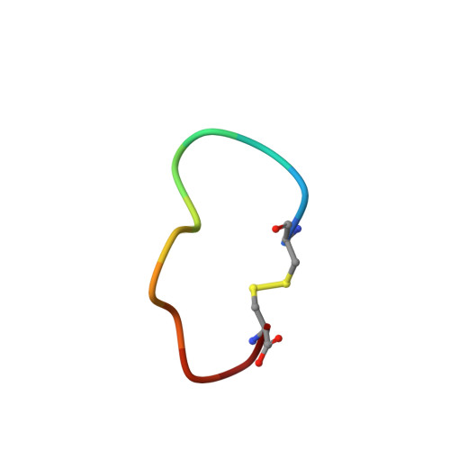
    Peptidic inhibitors of proteases are attracting increasing interest not only as drug candidates but also for studying the function and regulation mechanisms of these enzymes. Previously, we screened out a cyclic peptide inhibitor of human uPA [Formula: see text] and found that Ala substitution of P2 residue turns upain-1 to a substrate. To further investigate the effect of P2 residue on the peptide behavior transformation, we constructed upain-1-W3F, which has Phe replacement in the P2 position. We determined K D and K i of upain-1-W3F and found that upain-1-W3F might still exist as an inhibitor. Furthermore, the high-resolution crystal structure of upain-1-W3F·uPA reveals that upain-1-W3F indeed stays as an intact inhibitor bind to uPA. We thus propose that the P2 residue plays a nonnegligible role in the conversion of upain-1 to a substrate. These results also proposed a strategy to optimize the pharmacological properties of peptide-based drug candidates by hydrophobicity and steric hindrance. Abbreviations : uPA: urokinase-type plasminogen activator; SPD: serine protease domain; S1 pocket: specific substrate-binding pocket.


  • Organizational Affiliation

    College of Chemistry, Fuzhou University, Fuzhou, China.


Macromolecules
Find similar proteins by:  (by identity cutoff)  |  3D Structure
Entity ID: 1
MoleculeChains Sequence LengthOrganismDetailsImage
Urokinase-type plasminogen activatorA [auth U]253Homo sapiensMutation(s): 2 
Gene Names: PLAU
EC: 3.4.21.73
UniProt & NIH Common Fund Data Resources
Find proteins for P00749 (Homo sapiens)
Explore P00749 
Go to UniProtKB:  P00749
PHAROS:  P00749
GTEx:  ENSG00000122861 
Entity Groups  
Sequence Clusters30% Identity50% Identity70% Identity90% Identity95% Identity100% Identity
UniProt GroupP00749
Sequence Annotations
Expand
  • Reference Sequence

Find similar proteins by:  Sequence   |   3D Structure  

Entity ID: 2
MoleculeChains Sequence LengthOrganismDetailsImage
upain-1-W3FB [auth P]12synthetic constructMutation(s): 0 
Entity Groups  
Sequence Clusters30% Identity50% Identity70% Identity90% Identity95% Identity100% Identity
Sequence Annotations
Expand
  • Reference Sequence
Experimental Data & Validation

Experimental Data

  • Method: X-RAY DIFFRACTION
  • Resolution: 1.40 Å
  • R-Value Free: 0.203 
  • R-Value Work: 0.186 
  • R-Value Observed: 0.187 
  • Space Group: H 3
Unit Cell:
Length ( Å )Angle ( ˚ )
a = 121.292α = 90
b = 121.292β = 90
c = 43γ = 120
Software Package:
Software NamePurpose
PHENIXrefinement
PDB_EXTRACTdata extraction
HKL-3000data reduction
HKL-3000data scaling
PHENIXphasing

Structure Validation

View Full Validation Report


Entry History & Funding Information

Deposition Data


Funding OrganizationLocationGrant Number
National Natural Science Foundation of China (NSFC)China31400637
National Natural Science Foundation of China (NSFC)China31161130356
National Natural Science Foundation of China (NSFC)China31170707
National Natural Science Foundation of China (NSFC)China31370737

Revision History  (Full details and data files)

  • Version 1.0: 2020-02-19
    Type: Initial release
  • Version 1.1: 2020-05-13
    Changes: Database references
  • Version 1.2: 2024-01-24
    Changes: Data collection, Database references, Refinement description