6XUK

AbLIFT design 15 of Ab 1116NS19.9


Experimental Data Snapshot

  • Method: X-RAY DIFFRACTION
  • Resolution: 1.42 Å
  • R-Value Free: 0.185 
  • R-Value Work: 0.149 
  • R-Value Observed: 0.150 

wwPDB Validation   3D Report Full Report


This is version 1.4 of the entry. See complete history


Literature

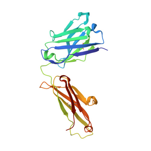
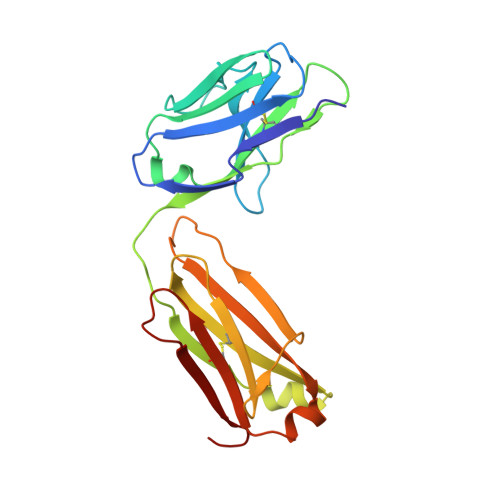
Biomolecular Recognition of the Glycan Neoantigen CA19-9 by Distinct Antibodies.

Borenstein-Katz, A.Warszawski, S.Amon, R.Eilon, M.Cohen-Dvashi, H.Leviatan Ben-Arye, S.Tasnima, N.Yu, H.Chen, X.Padler-Karavani, V.Fleishman, S.J.Diskin, R.

(2021) J Mol Biol 433: 167099-167099

  • DOI: https://doi.org/10.1016/j.jmb.2021.167099
  • Primary Citation of Related Structures:  
    6XTG, 6XUD, 6XUK, 6XUL, 6XUN

  • PubMed Abstract: 

    Glycans decorate the cell surface, secreted glycoproteins and glycolipids, and altered glycans are often found in cancers. Despite their high diagnostic and therapeutic potential, however, glycans are polar and flexible molecules that are quite challenging for the development and design of high-affinity binding antibodies. To understand the mechanisms by which glycan neoantigens are specifically recognized by antibodies, we analyze the biomolecular recognition of the tumor-associated carbohydrate antigen CA19-9 by two distinct antibodies using X-ray crystallography. Despite the potential plasticity of glycans and the very different antigen-binding surfaces presented by the antibodies, both structures reveal an essentially identical extended CA19-9 conformer, suggesting that this conformer's stability selects the antibodies. Starting from the bound structure of one of the antibodies, we use the AbLIFT computational algorithm to design a variant with seven core mutations in the variable domain's light-heavy chain interface that exhibits tenfold improved affinity for CA19-9. The results reveal strategies used by antibodies to specifically recognize glycan antigens and show how automated antibody-optimization methods may be used to enhance the clinical potential of existing antibodies.


  • Organizational Affiliation

    Department of Chemical and Structural Biology, Weizmann Institute of Science, 76100 Rehovot, Israel.


Macromolecules
Find similar proteins by:  (by identity cutoff)  |  3D Structure
Entity ID: 1
MoleculeChains Sequence LengthOrganismDetailsImage
Heavy chainA [auth H]222Mus musculusMutation(s): 0 
Entity Groups  
Sequence Clusters30% Identity50% Identity70% Identity90% Identity95% Identity100% Identity
Sequence Annotations
Expand
  • Reference Sequence
Find similar proteins by:  (by identity cutoff)  |  3D Structure
Entity ID: 2
MoleculeChains Sequence LengthOrganismDetailsImage
Light chainB [auth L]214Mus musculusMutation(s): 0 
Entity Groups  
Sequence Clusters30% Identity50% Identity70% Identity90% Identity95% Identity100% Identity
Sequence Annotations
Expand
  • Reference Sequence
Oligosaccharides

Help

Entity ID: 3
MoleculeChains Length2D Diagram Glycosylation3D Interactions
N-acetyl-alpha-neuraminic acid-(2-3)-beta-D-galactopyranose-(1-3)-[alpha-L-fucopyranose-(1-4)]2-acetamido-2-deoxy-beta-D-glucopyranoseC [auth A]4N/A
Glycosylation Resources
GlyTouCan:  G00053MO
GlyCosmos:  G00053MO
GlyGen:  G00053MO
Experimental Data & Validation

Experimental Data

  • Method: X-RAY DIFFRACTION
  • Resolution: 1.42 Å
  • R-Value Free: 0.185 
  • R-Value Work: 0.149 
  • R-Value Observed: 0.150 
  • Space Group: P 43 21 2
Unit Cell:
Length ( Å )Angle ( ˚ )
a = 64.936α = 90
b = 64.936β = 90
c = 244.109γ = 90
Software Package:
Software NamePurpose
PHENIXrefinement
xia2data scaling
PDB_EXTRACTdata extraction
xia2data reduction
PHASERphasing

Structure Validation

View Full Validation Report


Entry History 

Deposition Data

Revision History  (Full details and data files)

  • Version 1.0: 2021-02-03
    Type: Initial release
  • Version 1.1: 2021-08-11
    Changes: Advisory, Database references
  • Version 1.2: 2021-09-29
    Changes: Data collection, Database references
  • Version 1.3: 2023-03-08
    Changes: Advisory
  • Version 1.4: 2024-02-07
    Changes: Data collection, Refinement description