6XET

Tubulin-RB3_SLD in complex with compound 60c


Experimental Data Snapshot

  • Method: X-RAY DIFFRACTION
  • Resolution: 2.60 Å
  • R-Value Free: 0.231 
  • R-Value Work: 0.168 
  • R-Value Observed: 0.170 

wwPDB Validation   3D Report Full Report


Ligand Structure Quality Assessment 


This is version 1.2 of the entry. See complete history


Literature

Design, Synthesis, and Biological Evaluation of Stable Colchicine-Binding Site Tubulin Inhibitors 6-Aryl-2-benzoyl-pyridines as Potential Anticancer Agents.

Chen, H.Deng, S.Albadari, N.Yun, M.K.Zhang, S.Li, Y.Ma, D.Parke, D.N.Yang, L.Seagroves, T.N.White, S.W.Miller, D.D.Li, W.

(2021) J Med Chem 64: 12049-12074

  • DOI: https://doi.org/10.1021/acs.jmedchem.1c00715
  • Primary Citation of Related Structures:  
    6XER, 6XES, 6XET

  • PubMed Abstract: 

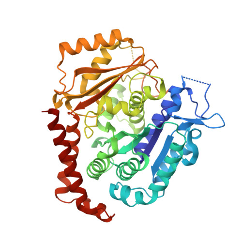
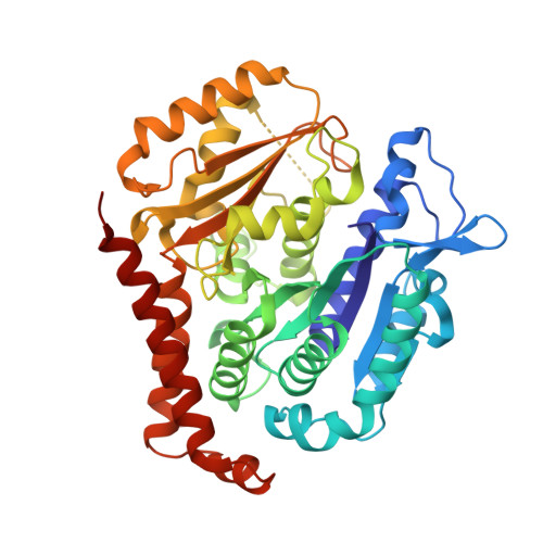
    We previously reported a potent tubulin inhibitor CH-2-77. In this study, we optimized the structure of CH-2-77 by blocking metabolically labile sites and synthesized a series of CH-2-77 analogues. Two compounds, 40a and 60c , preserved the potency while improving the metabolic stability over CH-2-77 by 3- to 4-fold (46.8 and 29.4 vs 10.8 min in human microsomes). We determined the high-resolution X-ray crystal structures of 40a (resolution 2.3 Å) and 60c (resolution 2.6 Å) in complex with tubulin and confirmed their direct binding at the colchicine-binding site. In vitro , 60c maintained its mode of action by inhibiting tubulin polymerization and was effective against P-glycoprotein-mediated multiple drug resistance and taxol resistance. In vivo , 60c exhibited a strong inhibitory effect on tumor growth and metastasis in a taxol-resistant A375/TxR xenograft model without obvious toxicity. Collectively, this work showed that 60c is a promising lead compound for further development as a potential anticancer agent.


  • Organizational Affiliation

    Department of Pharmaceutical Sciences, College of Pharmacy, University of Tennessee Health Science Center, Memphis, Tennessee 38163, United States.


Macromolecules
Find similar proteins by:  (by identity cutoff)  |  3D Structure
Entity ID: 1
MoleculeChains Sequence LengthOrganismDetailsImage
Tubulin alpha-1B chain
A, C
438Sus scrofaMutation(s): 0 
UniProt
Find proteins for Q2XVP4 (Sus scrofa)
Explore Q2XVP4 
Go to UniProtKB:  Q2XVP4
Entity Groups  
Sequence Clusters30% Identity50% Identity70% Identity90% Identity95% Identity100% Identity
UniProt GroupQ2XVP4
Sequence Annotations
Expand
  • Reference Sequence
Find similar proteins by:  (by identity cutoff)  |  3D Structure
Entity ID: 2
MoleculeChains Sequence LengthOrganismDetailsImage
Tubulin beta chain
B, D
433Sus scrofaMutation(s): 0 
UniProt
Find proteins for P02554 (Sus scrofa)
Explore P02554 
Go to UniProtKB:  P02554
Entity Groups  
Sequence Clusters30% Identity50% Identity70% Identity90% Identity95% Identity100% Identity
UniProt GroupP02554
Sequence Annotations
Expand
  • Reference Sequence
Find similar proteins by:  (by identity cutoff)  |  3D Structure
Entity ID: 3
MoleculeChains Sequence LengthOrganismDetailsImage
Stathmin-4143Rattus norvegicusMutation(s): 2 
Gene Names: Stmn4
UniProt
Find proteins for P63043 (Rattus norvegicus)
Explore P63043 
Go to UniProtKB:  P63043
Entity Groups  
Sequence Clusters30% Identity50% Identity70% Identity90% Identity95% Identity100% Identity
UniProt GroupP63043
Sequence Annotations
Expand
  • Reference Sequence
Small Molecules
Ligands 5 Unique
IDChains Name / Formula / InChI Key2D Diagram3D Interactions
GTP
Query on GTP

Download Ideal Coordinates CCD File 
F [auth A],
K [auth C],
M [auth D]
GUANOSINE-5'-TRIPHOSPHATE
C10 H16 N5 O14 P3
XKMLYUALXHKNFT-UUOKFMHZSA-N
GDP
Query on GDP

Download Ideal Coordinates CCD File 
H [auth B]GUANOSINE-5'-DIPHOSPHATE
C10 H15 N5 O11 P2
QGWNDRXFNXRZMB-UUOKFMHZSA-N
TU2 (Subject of Investigation/LOI)
Query on TU2

Download Ideal Coordinates CCD File 
I [auth B],
N [auth D]
[3-fluoro-6-(3-hydroxy-4-methylphenyl)pyridin-2-yl](3,4,5-trimethoxyphenyl)methanone
C22 H20 F N O5
JZHPUOFRNWNYQR-UHFFFAOYSA-N
SO4
Query on SO4

Download Ideal Coordinates CCD File 
G [auth A],
J [auth B],
O [auth D]
SULFATE ION
O4 S
QAOWNCQODCNURD-UHFFFAOYSA-L
MG
Query on MG

Download Ideal Coordinates CCD File 
L [auth C]MAGNESIUM ION
Mg
JLVVSXFLKOJNIY-UHFFFAOYSA-N
Experimental Data & Validation

Experimental Data

  • Method: X-RAY DIFFRACTION
  • Resolution: 2.60 Å
  • R-Value Free: 0.231 
  • R-Value Work: 0.168 
  • R-Value Observed: 0.170 
  • Space Group: P 21 21 21
Unit Cell:
Length ( Å )Angle ( ˚ )
a = 66.097α = 90
b = 126.938β = 90
c = 250.461γ = 90
Software Package:
Software NamePurpose
PHENIXrefinement
HKL-2000data reduction
HKL-2000data scaling
PHASERphasing

Structure Validation

View Full Validation Report


Ligand Structure Quality Assessment 


Entry History & Funding Information

Deposition Data


Funding OrganizationLocationGrant Number
National Institutes of Health/National Cancer Institute (NIH/NCI)United StatesR01CA148706

Revision History  (Full details and data files)

  • Version 1.0: 2021-08-25
    Type: Initial release
  • Version 1.1: 2021-09-08
    Changes: Database references
  • Version 1.2: 2023-10-18
    Changes: Data collection, Refinement description