6X9Z

De novo design of transmembrane beta-barrels


Experimental Data Snapshot

  • Method: X-RAY DIFFRACTION
  • Resolution: 2.05 Å
  • R-Value Free: 0.274 
  • R-Value Work: 0.260 
  • R-Value Observed: 0.261 

wwPDB Validation   3D Report Full Report


This is version 1.3 of the entry. See complete history


Literature

De novo design of transmembrane beta barrels.

Vorobieva, A.A.White, P.Liang, B.Horne, J.E.Bera, A.K.Chow, C.M.Gerben, S.Marx, S.Kang, A.Stiving, A.Q.Harvey, S.R.Marx, D.C.Khan, G.N.Fleming, K.G.Wysocki, V.H.Brockwell, D.J.Tamm, L.K.Radford, S.E.Baker, D.

(2021) Science 371

  • DOI: https://doi.org/10.1126/science.abc8182
  • Primary Citation of Related Structures:  
    6X1K, 6X9Z

  • PubMed Abstract: 

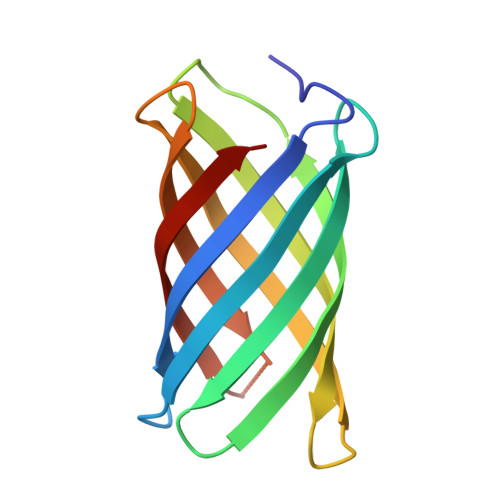
    Transmembrane β-barrel proteins (TMBs) are of great interest for single-molecule analytical technologies because they can spontaneously fold and insert into membranes and form stable pores, but the range of pore properties that can be achieved by repurposing natural TMBs is limited. We leverage the power of de novo computational design coupled with a "hypothesis, design, and test" approach to determine TMB design principles, notably, the importance of negative design to slow β-sheet assembly. We design new eight-stranded TMBs, with no homology to known TMBs, that insert and fold reversibly into synthetic lipid membranes and have nuclear magnetic resonance and x-ray crystal structures very similar to the computational models. These advances should enable the custom design of pores for a wide range of applications.


  • Organizational Affiliation

    Department of Biochemistry, University of Washington, Seattle, WA 98195, USA.


Macromolecules
Find similar proteins by:  (by identity cutoff)  |  3D Structure
Entity ID: 1
MoleculeChains Sequence LengthOrganismDetailsImage
Transmembrane beta-barrels124synthetic constructMutation(s): 0 
Entity Groups  
Sequence Clusters30% Identity50% Identity70% Identity90% Identity95% Identity100% Identity
Sequence Annotations
Expand
  • Reference Sequence
Experimental Data & Validation

Experimental Data

  • Method: X-RAY DIFFRACTION
  • Resolution: 2.05 Å
  • R-Value Free: 0.274 
  • R-Value Work: 0.260 
  • R-Value Observed: 0.261 
  • Space Group: H 3
Unit Cell:
Length ( Å )Angle ( ˚ )
a = 51.083α = 90
b = 51.083β = 90
c = 116.71γ = 120
Software Package:
Software NamePurpose
PHENIXrefinement
XDSdata reduction
XDSdata scaling
PHASERphasing

Structure Validation

View Full Validation Report


Entry History & Funding Information

Deposition Data


Funding OrganizationLocationGrant Number
Howard Hughes Medical Institute (HHMI)United States--

Revision History  (Full details and data files)

  • Version 1.0: 2021-02-17
    Type: Initial release
  • Version 1.1: 2021-03-03
    Changes: Database references
  • Version 1.2: 2024-03-06
    Changes: Data collection, Database references
  • Version 1.3: 2024-04-03
    Changes: Refinement description