6X6H

Structure of Shiga toxin 2 with a C-terminal peptide of ribosomal P stalk proteins


Experimental Data Snapshot

  • Method: X-RAY DIFFRACTION
  • Resolution: 1.88 Å
  • R-Value Free: 0.206 
  • R-Value Work: 0.168 
  • R-Value Observed: 0.170 

wwPDB Validation   3D Report Full Report


This is version 2.2 of the entry. See complete history


Literature

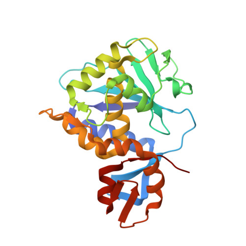
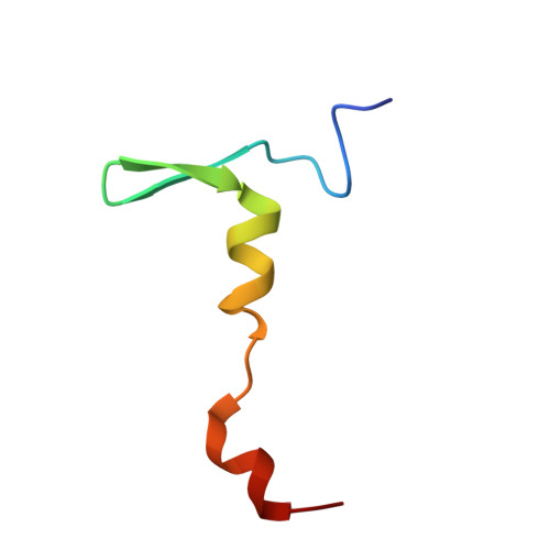
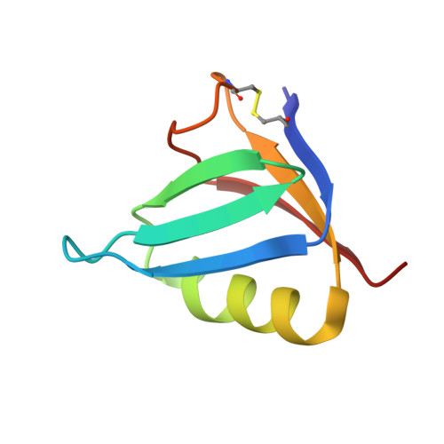
Structural basis for the interaction of Shiga toxin 2a with a C-terminal peptide of ribosomal P stalk proteins.

Rudolph, M.J.Davis, S.A.Tumer, N.E.Li, X.P.

(2020) J Biol Chem 295: 15588-15596

  • DOI: https://doi.org/10.1074/jbc.AC120.015070
  • Primary Citation of Related Structures:  
    6X6H

  • PubMed Abstract: 

    The principal virulence factor of human pathogenic enterohemorrhagic Escherichia coli is Shiga toxin (Stx). Shiga toxin 2a (Stx2a) is the subtype most commonly associated with severe disease outcomes such as hemorrhagic colitis and hemolytic uremic syndrome. The catalytic A1 subunit (Stx2A1) binds to the conserved elongation factor binding C-terminal domain (CTD) of ribosomal P stalk proteins to inhibit translation. Stx2a holotoxin also binds to the CTD of P stalk proteins because the ribosome-binding site is exposed. We show here that Stx2a binds to an 11-mer peptide (P11) mimicking the CTD of P stalk proteins with low micromolar affinity. We cocrystallized Stx2a with P11 and defined their interactions by X-ray crystallography. We found that the last six residues of P11 inserted into a shallow pocket on Stx2A1 and interacted with Arg-172, Arg-176, and Arg-179, which were previously shown to be critical for binding of Stx2A1 to the ribosome. Stx2a formed a distinct P11-binding mode within a different surface pocket relative to ricin toxin A subunit and trichosanthin, suggesting different ribosome recognition mechanisms for each ribosome inactivating protein (RIP). The binding mode of Stx2a to P11 is also conserved among the different Stx subtypes. Furthermore, the P stalk protein CTD is flexible and adopts distinct orientations and interaction modes depending on the structural differences between the RIPs. Structural characterization of the Stx2a-ribosome complex is important for understanding the role of the stalk in toxin recruitment to the sarcin/ricin loop and may provide a new target for inhibitor discovery.


  • Organizational Affiliation

    New York Structural Biology Center, New York, New York, USA.


Macromolecules
Find similar proteins by:  (by identity cutoff)  |  3D Structure
Entity ID: 1
MoleculeChains Sequence LengthOrganismDetailsImage
rRNA N-glycosylaseA [auth A1]242Escherichia coliMutation(s): 0 
Gene Names: 
EC: 3.2.2.22
UniProt
Find proteins for A9ZMR8 (Escherichia coli)
Explore A9ZMR8 
Go to UniProtKB:  A9ZMR8
Entity Groups  
Sequence Clusters30% Identity50% Identity70% Identity90% Identity95% Identity100% Identity
UniProt GroupA9ZMR8
Sequence Annotations
Expand
  • Reference Sequence
Find similar proteins by:  (by identity cutoff)  |  3D Structure
Entity ID: 2
MoleculeChains Sequence LengthOrganismDetailsImage
rRNA N-glycosylaseB [auth A2]40Escherichia coliMutation(s): 0 
Gene Names: 
EC: 3.2.2.22
UniProt
Find proteins for A9ZMR8 (Escherichia coli)
Explore A9ZMR8 
Go to UniProtKB:  A9ZMR8
Entity Groups  
Sequence Clusters30% Identity50% Identity70% Identity90% Identity95% Identity100% Identity
UniProt GroupA9ZMR8
Sequence Annotations
Expand
  • Reference Sequence
Find similar proteins by:  (by identity cutoff)  |  3D Structure
Entity ID: 3
MoleculeChains Sequence LengthOrganismDetailsImage
Shiga toxin 2 B subunitC [auth B],
D [auth C],
E [auth D],
F [auth E],
G [auth F]
70Escherichia coliMutation(s): 0 
Gene Names: 
UniProt
Find proteins for Q7DJJ2 (Escherichia coli)
Explore Q7DJJ2 
Go to UniProtKB:  Q7DJJ2
Entity Groups  
Sequence Clusters30% Identity50% Identity70% Identity90% Identity95% Identity100% Identity
UniProt GroupQ7DJJ2
Sequence Annotations
Expand
  • Reference Sequence

Find similar proteins by:  Sequence   |   3D Structure  

Entity ID: 4
MoleculeChains Sequence LengthOrganismDetailsImage
P11 peptideH [auth P]6Escherichia coliMutation(s): 0 
Sequence Annotations
Expand
  • Reference Sequence
Small Molecules
Ligands 3 Unique
IDChains Name / Formula / InChI Key2D Diagram3D Interactions
EDO
Query on EDO

Download Ideal Coordinates CCD File 
M [auth B],
O [auth C],
R [auth D],
S [auth F],
T [auth F]
1,2-ETHANEDIOL
C2 H6 O2
LYCAIKOWRPUZTN-UHFFFAOYSA-N
ACY
Query on ACY

Download Ideal Coordinates CCD File 
N [auth B],
U [auth F]
ACETIC ACID
C2 H4 O2
QTBSBXVTEAMEQO-UHFFFAOYSA-N
CL
Query on CL

Download Ideal Coordinates CCD File 
I [auth A1]
J [auth A1]
K [auth A1]
L [auth A2]
P [auth D]
I [auth A1],
J [auth A1],
K [auth A1],
L [auth A2],
P [auth D],
Q [auth D]
CHLORIDE ION
Cl
VEXZGXHMUGYJMC-UHFFFAOYSA-M
Experimental Data & Validation

Experimental Data

  • Method: X-RAY DIFFRACTION
  • Resolution: 1.88 Å
  • R-Value Free: 0.206 
  • R-Value Work: 0.168 
  • R-Value Observed: 0.170 
  • Space Group: P 2 21 21
Unit Cell:
Length ( Å )Angle ( ˚ )
a = 82.148α = 90
b = 85.226β = 90
c = 98.841γ = 90
Software Package:
Software NamePurpose
DENZOdata reduction
SCALEPACKdata scaling
PHASERphasing
PHENIXrefinement
PDB_EXTRACTdata extraction

Structure Validation

View Full Validation Report


Entry History 

Deposition Data

Revision History  (Full details and data files)

  • Version 1.0: 2020-09-09
    Type: Initial release
  • Version 2.0: 2020-09-16
    Changes: Advisory, Atomic model, Data collection, Database references, Derived calculations, Polymer sequence, Structure summary
  • Version 2.1: 2020-11-25
    Changes: Database references
  • Version 2.2: 2023-10-18
    Changes: Data collection, Database references, Refinement description