6X5Z

Bovine Cardiac Myosin in Complex with Chicken Skeletal Actin and Human Cardiac Tropomyosin in the Rigor State


Experimental Data Snapshot

  • Method: ELECTRON MICROSCOPY
  • Resolution: 4.24 Å
  • Aggregation State: HELICAL ARRAY 
  • Reconstruction Method: HELICAL 

wwPDB Validation   3D Report Full Report


This is version 1.3 of the entry. See complete history


Literature

Cryo-EM and Molecular Docking Shows Myosin Loop 4 Contacts Actin and Tropomyosin on Thin Filaments.

Doran, M.H.Pavadai, E.Rynkiewicz, M.J.Walklate, J.Bullitt, E.Moore, J.R.Regnier, M.Geeves, M.A.Lehman, W.

(2020) Biophys J 119: 821-830

  • DOI: https://doi.org/10.1016/j.bpj.2020.07.006
  • Primary Citation of Related Structures:  
    6X5Z

  • PubMed Abstract: 

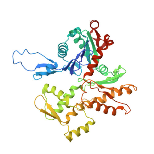
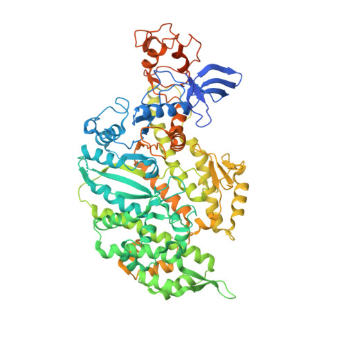
    The motor protein myosin drives muscle and nonmuscle motility by binding to and moving along actin of thin filaments. Myosin binding to actin also modulates interactions of the regulatory protein, tropomyosin, on thin filaments, and conversely tropomyosin affects myosin binding to actin. Insight into this reciprocity will facilitate a molecular level elucidation of tropomyosin regulation of myosin interaction with actin in muscle contraction, and in turn, promote better understanding of nonmuscle cell motility. Indeed, experimental approaches such as fiber diffraction, cryoelectron microscopy, and three-dimensional reconstruction have long been used to define regulatory interaction of tropomyosin and myosin on actin at a structural level. However, their limited resolution has not proven sufficient to determine tropomyosin and myosin contacts at an atomic-level and thus to fully substantiate possible functional contributions. To overcome this deficiency, we have followed a hybrid approach by performing new cryogenic electron microscopy reconstruction of myosin-S1-decorated F-actin-tropomyosin together with atomic scale protein-protein docking of tropomyosin to the EM models. Here, cryo-EM data were derived from filaments reconstituted with α1-actin, cardiac αα-tropomyosin, and masseter muscle β-myosin complexes; masseter myosin, which shares sequence identity with β-cardiac myosin-heavy chain, was used because of its stability in vitro. The data were used to build an atomic model of the tropomyosin cable that fits onto the actin filament between the tip of the myosin head and a cleft on the innermost edge of actin subunits. The docking and atomic scale fitting showed multiple discrete interactions of myosin loop 4 and acidic residues on successive 39-42 residue-long tropomyosin pseudorepeats. The contacts between S1 and tropomyosin on actin appear to compete with and displace ones normally found between actin and tropomyosin on myosin-free thin filaments in relaxed muscle, thus restructuring the filament during myosin-induced activation.


  • Organizational Affiliation

    Department of Physiology & Biophysics, Boston University School of Medicine, Boston, Massachusetts.


Macromolecules
Find similar proteins by:  (by identity cutoff)  |  3D Structure
Entity ID: 1
MoleculeChains Sequence LengthOrganismDetailsImage
Actin, alpha skeletal muscleA [auth B],
E [auth A],
G [auth C]
377Gallus gallusMutation(s): 0 
UniProt
Find proteins for P68139 (Gallus gallus)
Explore P68139 
Go to UniProtKB:  P68139
Entity Groups  
Sequence Clusters30% Identity50% Identity70% Identity90% Identity95% Identity100% Identity
UniProt GroupP68139
Sequence Annotations
Expand
  • Reference Sequence
Find similar proteins by:  (by identity cutoff)  |  3D Structure
Entity ID: 2
MoleculeChains Sequence LengthOrganismDetailsImage
Tropomyosin alpha-1 chainB [auth O],
D [auth P]
284Homo sapiensMutation(s): 0 
Gene Names: TPM1C15orf13TMSA
UniProt & NIH Common Fund Data Resources
Find proteins for P09493 (Homo sapiens)
Explore P09493 
Go to UniProtKB:  P09493
PHAROS:  P09493
GTEx:  ENSG00000140416 
Entity Groups  
Sequence Clusters30% Identity50% Identity70% Identity90% Identity95% Identity100% Identity
UniProt GroupP09493
Sequence Annotations
Expand
  • Reference Sequence
Find similar proteins by:  (by identity cutoff)  |  3D Structure
Entity ID: 3
MoleculeChains Sequence LengthOrganismDetailsImage
Myosin-7C [auth G],
F [auth D],
H [auth J]
850Bos taurusMutation(s): 0 
UniProt
Find proteins for Q9BE39 (Bos taurus)
Explore Q9BE39 
Go to UniProtKB:  Q9BE39
Entity Groups  
Sequence Clusters30% Identity50% Identity70% Identity90% Identity95% Identity100% Identity
UniProt GroupQ9BE39
Sequence Annotations
Expand
  • Reference Sequence
Experimental Data & Validation

Experimental Data

  • Method: ELECTRON MICROSCOPY
  • Resolution: 4.24 Å
  • Aggregation State: HELICAL ARRAY 
  • Reconstruction Method: HELICAL 
EM Software:
TaskSoftware PackageVersion
RECONSTRUCTIONRELION

Structure Validation

View Full Validation Report


Entry History & Funding Information

Deposition Data


Funding OrganizationLocationGrant Number
National Institutes of Health/National Heart, Lung, and Blood Institute (NIH/NHLBI)United StatesR01HL036153
National Institutes of Health/National Heart, Lung, and Blood Institute (NIH/NHLBI)United StatesR01HL123774
National Institutes of Health/National Institute of General Medical Sciences (NIH/NIGMS)United StatesS10RR25434
National Institutes of Health/National Institute of General Medical Sciences (NIH/NIGMS)United StatesU24 GM129541
National Institutes of Health/National Institute of General Medical Sciences (NIH/NIGMS)United StatesR01GM029090
National Institutes of Health/National Institute of General Medical Sciences (NIH/NIGMS)United StatesU24GM116787
Horizon 2020 Research and Innovation ProgrammeEuropean Union777204 SILICOFCM

Revision History  (Full details and data files)

  • Version 1.0: 2020-07-22
    Type: Initial release
  • Version 1.1: 2020-08-12
    Changes: Database references
  • Version 1.2: 2020-09-02
    Changes: Database references
  • Version 1.3: 2024-03-06
    Changes: Data collection, Database references, Refinement description