6WZS

Fusibacterium ulcerans ZTP riboswitch bound to m-1-pyridinyl AICA


Experimental Data Snapshot

  • Method: X-RAY DIFFRACTION
  • Resolution: 3.23 Å
  • R-Value Free: 0.259 
  • R-Value Work: 0.224 
  • R-Value Observed: 0.227 

wwPDB Validation   3D Report Full Report


Ligand Structure Quality Assessment 


This is version 1.1 of the entry. See complete history


Literature

Parallel Discovery Strategies Provide a Basis for Riboswitch Ligand Design.

Tran, B.Pichling, P.Tenney, L.Connelly, C.M.Moon, M.H.Ferre-D'Amare, A.R.Schneekloth Jr., J.S.Jones, C.P.

(2020) Cell Chem Biol 27: 1241-1249.e4

  • DOI: https://doi.org/10.1016/j.chembiol.2020.07.021
  • Primary Citation of Related Structures:  
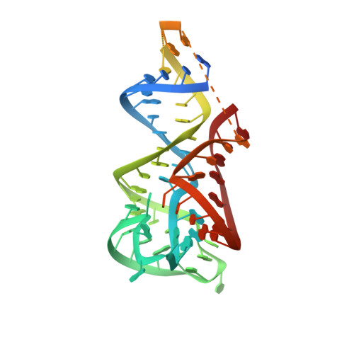
    6WZR, 6WZS

  • PubMed Abstract: 

    Riboswitches are mRNA domains that make gene-regulatory decisions upon binding their cognate ligands. Bacterial riboswitches that specifically recognize 5-aminoimidazole-4-carboxamide riboside 5'-monophosphate (ZMP) and 5'-triphosphate (ZTP) regulate genes involved in folate and purine metabolism. Now, we have developed synthetic ligands targeting ZTP riboswitches by replacing the sugar-phosphate moiety of ZMP with various functional groups, including simple heterocycles. Despite losing hydrogen bonds from ZMP, these analogs bind ZTP riboswitches with similar affinities as the natural ligand, and activate transcription more strongly than ZMP in vitro. The most active ligand stimulates gene expression ∼3 times more than ZMP in a live Escherichia coli reporter. Co-crystal structures of the Fusobacterium ulcerans ZTP riboswitch bound to synthetic ligands suggest stacking of their pyridine moieties on a conserved RNA nucleobase primarily determines their higher activity. Altogether, these findings guide future design of improved riboswitch activators and yield insights into how RNA-targeted ligand discovery may proceed.


  • Organizational Affiliation

    Biochemistry and Biophysics Center, National Heart, Lung, and Blood Institute, National Institutes of Health, Bethesda, MD, USA.


Macromolecules
Find similar nucleic acids by:  (by identity cutoff)  |  3D Structure
Entity ID: 1
MoleculeChains LengthOrganismImage
Fusibacterium ulcerans ZTP riboswitchA [auth B],
B [auth A]
75Fusobacterium ulcerans
Sequence Annotations
Expand
  • Reference Sequence
Small Molecules
Ligands 3 Unique
IDChains Name / Formula / InChI Key2D Diagram3D Interactions
UG4 (Subject of Investigation/LOI)
Query on UG4

Download Ideal Coordinates CCD File 
I [auth B],
P [auth A]
5-amino-1-(pyridin-3-yl)-1H-imidazole-4-carboxamide
C9 H9 N5 O
WZVCHXNAUQIDJM-UHFFFAOYSA-N
K
Query on K

Download Ideal Coordinates CCD File 
C [auth B],
D [auth B],
J [auth A],
K [auth A]
POTASSIUM ION
K
NPYPAHLBTDXSSS-UHFFFAOYSA-N
MG
Query on MG

Download Ideal Coordinates CCD File 
E [auth B]
F [auth B]
G [auth B]
H [auth B]
L [auth A]
E [auth B],
F [auth B],
G [auth B],
H [auth B],
L [auth A],
M [auth A],
N [auth A],
O [auth A]
MAGNESIUM ION
Mg
JLVVSXFLKOJNIY-UHFFFAOYSA-N
Experimental Data & Validation

Experimental Data

  • Method: X-RAY DIFFRACTION
  • Resolution: 3.23 Å
  • R-Value Free: 0.259 
  • R-Value Work: 0.224 
  • R-Value Observed: 0.227 
  • Space Group: P 32 2 1
Unit Cell:
Length ( Å )Angle ( ˚ )
a = 90.284α = 90
b = 90.284β = 90
c = 121.391γ = 120
Software Package:
Software NamePurpose
PHENIXrefinement
HKL-2000data scaling
PHASERphasing
PDB_EXTRACTdata extraction
HKL-2000data reduction

Structure Validation

View Full Validation Report


Ligand Structure Quality Assessment 


Entry History 

Deposition Data

Revision History  (Full details and data files)

  • Version 1.0: 2021-03-24
    Type: Initial release
  • Version 1.1: 2023-10-18
    Changes: Data collection, Database references, Refinement description