6WZN

Crystal Structure of a Fluorescent Single Chain Fv Chimera


Experimental Data Snapshot

  • Method: X-RAY DIFFRACTION
  • Resolution: 2.50 Å
  • R-Value Free: 0.211 
  • R-Value Work: 0.163 
  • R-Value Observed: 0.167 

wwPDB Validation   3D Report Full Report


This is version 2.0 of the entry. See complete history


Literature

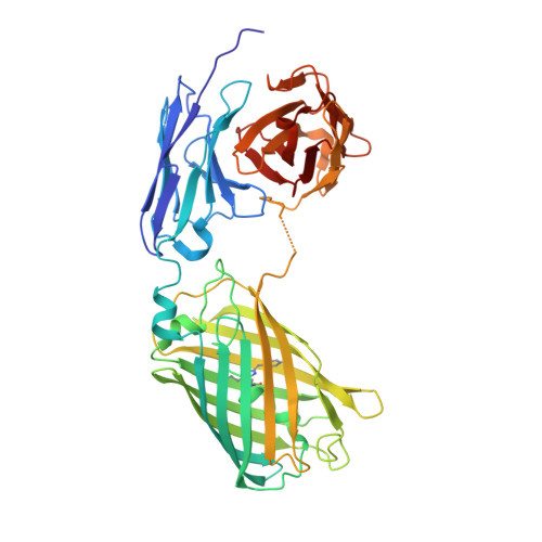
Construction, characterization and crystal structure of a fluorescent single-chain Fv chimera.

Velappan, N.Close, D.Hung, L.W.Naranjo, L.Hemez, C.DeVore, N.McCullough, D.K.Lillo, A.M.Waldo, G.S.Bradbury, A.R.M.

(2021) Protein Eng Des Sel 34

  • DOI: https://doi.org/10.1093/protein/gzaa029
  • Primary Citation of Related Structures:  
    6WZN

  • PubMed Abstract: 

    In vitro display technologies based on phage and yeast have a successful history of selecting single-chain variable fragment (scFv) antibodies against various targets. However, single-chain antibodies are often unstable and poorly expressed in Escherichia coli. Here, we explore the feasibility of converting scFv antibodies to an intrinsically fluorescent format by inserting the monomeric, stable fluorescent protein named thermal green, between the light- and heavy-chain variable regions. Our results show that the scTGP format maintains the affinity and specificity of the antibodies, improves expression levels, allows one-step fluorescent assay for detection of binding and is a suitable reagent for epitope binning. We also report the crystal structure of an scTGP construct that recognizes phosphorylated tyrosine on FcεR1 receptor of the allergy pathway.


  • Organizational Affiliation

    Bioscience Division, Los Alamos National Laboratory, Los Alamos, NM 87545, USA.


Macromolecules
Find similar proteins by:  (by identity cutoff)  |  3D Structure
Entity ID: 1
MoleculeChains Sequence LengthOrganismDetailsImage
Fluorescent Single Chain Fv Chimera463synthetic constructMutation(s): 0 
Entity Groups  
Sequence Clusters30% Identity50% Identity70% Identity90% Identity95% Identity100% Identity
Sequence Annotations
Expand
  • Reference Sequence
Small Molecules
Modified Residues  1 Unique
IDChains TypeFormula2D DiagramParent
CRQ
Query on CRQ
A
L-PEPTIDE LINKINGC16 H16 N4 O5GLN, TYR, GLY
Experimental Data & Validation

Experimental Data

  • Method: X-RAY DIFFRACTION
  • Resolution: 2.50 Å
  • R-Value Free: 0.211 
  • R-Value Work: 0.163 
  • R-Value Observed: 0.167 
  • Space Group: P 41 21 2
Unit Cell:
Length ( Å )Angle ( ˚ )
a = 103.961α = 90
b = 103.961β = 90
c = 122.74γ = 90
Software Package:
Software NamePurpose
HKL-2000data scaling
PHENIXrefinement
PDB_EXTRACTdata extraction
HKL-2000data reduction
PHENIXphasing

Structure Validation

View Full Validation Report


Entry History & Funding Information

Deposition Data


Funding OrganizationLocationGrant Number
Department of Energy (DOE, United States)United StatesP50GM085273

Revision History  (Full details and data files)

  • Version 1.0: 2021-01-27
    Type: Initial release
  • Version 1.1: 2021-02-10
    Changes: Source and taxonomy
  • Version 1.2: 2021-03-03
    Changes: Database references
  • Version 1.3: 2023-10-18
    Changes: Data collection, Database references, Refinement description
  • Version 2.0: 2023-11-15
    Changes: Atomic model, Data collection, Derived calculations