6WPL

Structure of Cytochrome P450tcu


Experimental Data Snapshot

  • Method: X-RAY DIFFRACTION
  • Resolution: 2.10 Å
  • R-Value Free: 0.247 
  • R-Value Work: 0.193 
  • R-Value Observed: 0.196 

wwPDB Validation   3D Report Full Report


Ligand Structure Quality Assessment 


This is version 1.2 of the entry. See complete history


Literature

Unexpected Differences between Two Closely Related Bacterial P450 Camphor Monooxygenases.

Murarka, V.C.Batabyal, D.Amaya, J.A.Sevrioukova, I.F.Poulos, T.L.

(2020) Biochemistry 59: 2743-2750

  • DOI: https://doi.org/10.1021/acs.biochem.0c00366
  • Primary Citation of Related Structures:  
    6WPL

  • PubMed Abstract: 

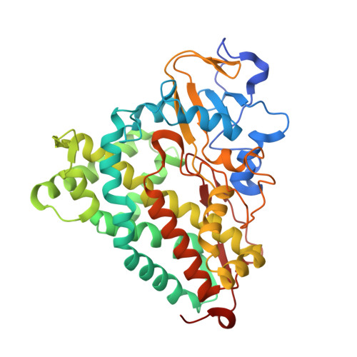
    The bacterial cytochrome P450cam catalyzes the oxidation of camphor to 5- exo -hydroxycamphor as the first step in the oxidative assimilation of camphor as a carbon/energy source. CYP101D1 is another bacterial P450 that catalyzes the same reaction. A third P450 (P450tcu) has recently been discovered that has ≈86% sequence identity to P450cam as well as very similar enzymatic properties. P450tcu, however, exhibits three unusual features not found in P450cam. First, we observe product in at least two orientations in the X-ray structure that indicates that, unlike the case for P450cam, X-ray-generated reducing equivalents can drive substrate hydroxylation in crystallo . We postulate, on the basis of molecular dynamics simulations, that greater flexibility in P450tcu enables easier access of protons to the active site and, together with X-ray driven reduction, results in O 2 activation and substrate hydroxylation. Second, the characteristic low-spin to high-spin transition when camphor binds occurs immediately with P450cam but is very slow in P450tcu. Third, isothermal titration calorimetry shows that in P450cam substrate binding is entropically driven with a Δ H of >0 while in P450tcu with a Δ H of <0 with a more modest change in - T Δ S . These results indicate that despite nearly identical structures and enzymatic properties, these two P450s exhibit quite different properties most likely related to differences in conformational dynamics.


  • Organizational Affiliation

    Departments of Molecular Biology and Biochemistry, Pharmaceutical Sciences, and Chemistry, University of California, Irvine, California 92697-3900, United States.


Macromolecules
Find similar proteins by:  (by identity cutoff)  |  3D Structure
Entity ID: 1
MoleculeChains Sequence LengthOrganismDetailsImage
Cytochrome P-450cam, subunit of camphor 5-monooxygenase system415Pseudomonas sp. TCU-HL1Mutation(s): 0 
Gene Names: THL1_4171
UniProt
Find proteins for A0A1B3EAY9 (Pseudomonas sp. TCU-HL1)
Explore A0A1B3EAY9 
Go to UniProtKB:  A0A1B3EAY9
Entity Groups  
Sequence Clusters30% Identity50% Identity70% Identity90% Identity95% Identity100% Identity
UniProt GroupA0A1B3EAY9
Sequence Annotations
Expand
  • Reference Sequence
Small Molecules
Binding Affinity Annotations 
IDSourceBinding Affinity
CAH Binding MOAD:  6WPL Kd: 702 (nM) from 1 assay(s)
Experimental Data & Validation

Experimental Data

  • Method: X-RAY DIFFRACTION
  • Resolution: 2.10 Å
  • R-Value Free: 0.247 
  • R-Value Work: 0.193 
  • R-Value Observed: 0.196 
  • Space Group: P 21 21 21
Unit Cell:
Length ( Å )Angle ( ˚ )
a = 35.42α = 90
b = 56.993β = 90
c = 189.373γ = 90
Software Package:
Software NamePurpose
PHENIXrefinement
XDSdata reduction
Aimlessdata scaling
PHASERphasing

Structure Validation

View Full Validation Report


Ligand Structure Quality Assessment 


Entry History & Funding Information

Deposition Data


Funding OrganizationLocationGrant Number
National Institutes of Health/Office of the DirectorUnited StatesGM131920
National Institutes of Health/Office of the DirectorUnited StatesES025767

Revision History  (Full details and data files)

  • Version 1.0: 2020-07-01
    Type: Initial release
  • Version 1.1: 2020-08-26
    Changes: Database references, Derived calculations
  • Version 1.2: 2023-10-18
    Changes: Data collection, Database references, Refinement description