6WNB

Structure of the Rieske non-heme iron oxygenase SxtT with dideoxysaxitoxin bound


Experimental Data Snapshot

  • Method: X-RAY DIFFRACTION
  • Resolution: 2.10 Å
  • R-Value Free: 0.207 
  • R-Value Work: 0.168 
  • R-Value Observed: 0.170 

wwPDB Validation   3D Report Full Report


Ligand Structure Quality Assessment 


This is version 1.2 of the entry. See complete history


Literature

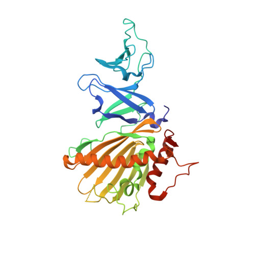
Structural basis for divergent C-H hydroxylation selectivity in two Rieske oxygenases.

Lukowski, A.L.Liu, J.Bridwell-Rabb, J.Narayan, A.R.H.

(2020) Nat Commun 11: 2991-2991

  • DOI: https://doi.org/10.1038/s41467-020-16729-0
  • Primary Citation of Related Structures:  
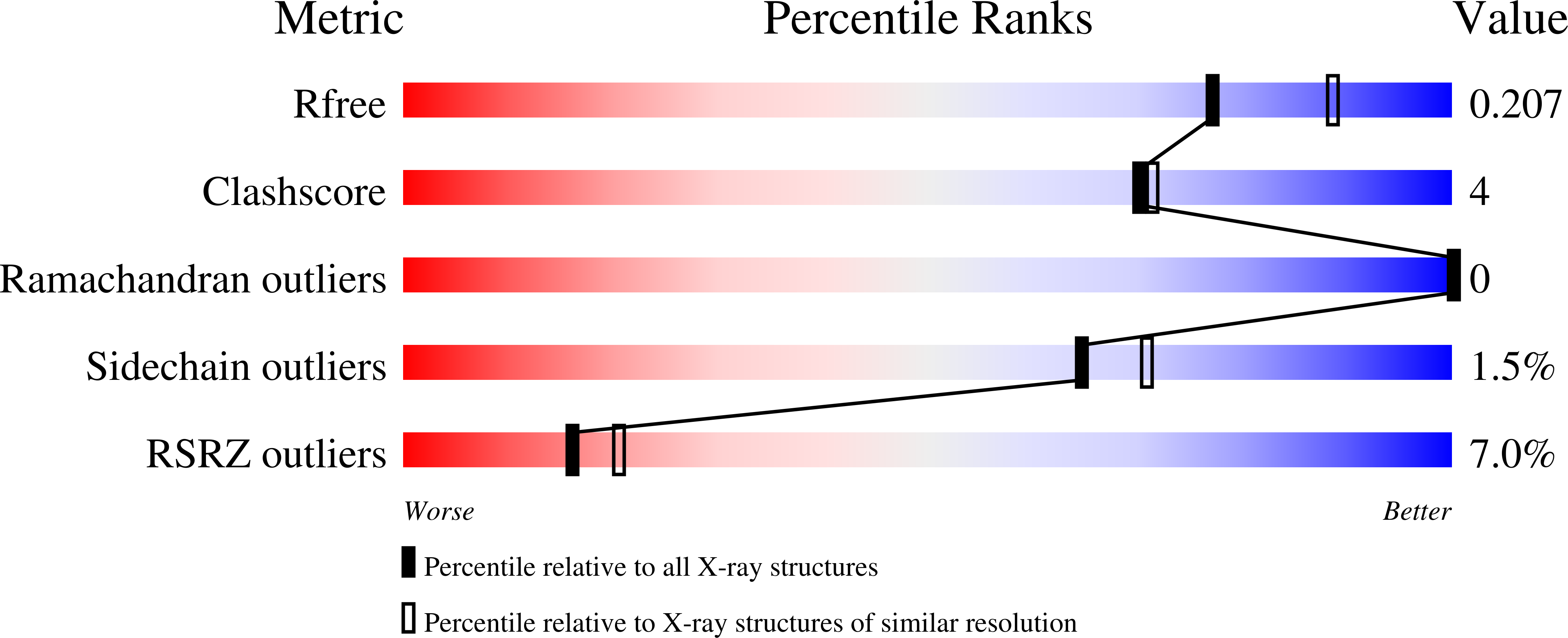
    6WN3, 6WNB, 6WNC, 6WND

  • PubMed Abstract: 

    Biocatalysts that perform C-H hydroxylation exhibit exceptional substrate specificity and site-selectivity, often through the use of high valent oxidants to activate these inert bonds. Rieske oxygenases are examples of enzymes with the ability to perform precise mono- or dioxygenation reactions on a variety of substrates. Understanding the structural features of Rieske oxygenases responsible for control over selectivity is essential to enable the development of this class of enzymes for biocatalytic applications. Decades of research has illuminated the critical features common to Rieske oxygenases, however, structural information for enzymes that functionalize diverse scaffolds is limited. Here, we report the structures of two Rieske monooxygenases involved in the biosynthesis of paralytic shellfish toxins (PSTs), SxtT and GxtA, adding to the short list of structurally characterized Rieske oxygenases. Based on these structures, substrate-bound structures, and mutagenesis experiments, we implicate specific residues in substrate positioning and the divergent reaction selectivity observed in these two enzymes.


  • Organizational Affiliation

    Program in Chemical Biology, University of Michigan, Ann Arbor, MI, USA.


Macromolecules
Find similar proteins by:  (by identity cutoff)  |  3D Structure
Entity ID: 1
MoleculeChains Sequence LengthOrganismDetailsImage
SxtT
A, B, C
334Microseira wolleiMutation(s): 0 
Gene Names: sxtT
UniProt
Find proteins for C3RVQ0 (Microseira wollei)
Explore C3RVQ0 
Go to UniProtKB:  C3RVQ0
Entity Groups  
Sequence Clusters30% Identity50% Identity70% Identity90% Identity95% Identity100% Identity
UniProt GroupC3RVQ0
Sequence Annotations
Expand
  • Reference Sequence
Small Molecules
Ligands 5 Unique
IDChains Name / Formula / InChI Key2D Diagram3D Interactions
U5A (Subject of Investigation/LOI)
Query on U5A

Download Ideal Coordinates CCD File 
BA [auth C],
G [auth A],
R [auth B]
[(2Z,3aS,4R,6Z,10aR)-2,6-diiminooctahydro-1H,8H-pyrrolo[1,2-c]purin-4-yl]methyl carbamate
C10 H17 N7 O2
WSPXQONARWJOKG-JFWOZONXSA-N
FES (Subject of Investigation/LOI)
Query on FES

Download Ideal Coordinates CCD File 
D [auth A],
N [auth B],
Y [auth C]
FE2/S2 (INORGANIC) CLUSTER
Fe2 S2
NIXDOXVAJZFRNF-UHFFFAOYSA-N
SO4
Query on SO4

Download Ideal Coordinates CCD File 
DA [auth C]
EA [auth C]
FA [auth C]
I [auth A]
J [auth A]
DA [auth C],
EA [auth C],
FA [auth C],
I [auth A],
J [auth A],
K [auth A],
L [auth A],
M [auth A],
T [auth B],
U [auth B],
V [auth B],
W [auth B],
X [auth B]
SULFATE ION
O4 S
QAOWNCQODCNURD-UHFFFAOYSA-L
GOL
Query on GOL

Download Ideal Coordinates CCD File 
AA [auth C]
E [auth A]
F [auth A]
O [auth B]
P [auth B]
AA [auth C],
E [auth A],
F [auth A],
O [auth B],
P [auth B],
Q [auth B],
Z [auth C]
GLYCEROL
C3 H8 O3
PEDCQBHIVMGVHV-UHFFFAOYSA-N
FE (Subject of Investigation/LOI)
Query on FE

Download Ideal Coordinates CCD File 
CA [auth C],
H [auth A],
S [auth B]
FE (III) ION
Fe
VTLYFUHAOXGGBS-UHFFFAOYSA-N
Experimental Data & Validation

Experimental Data

  • Method: X-RAY DIFFRACTION
  • Resolution: 2.10 Å
  • R-Value Free: 0.207 
  • R-Value Work: 0.168 
  • R-Value Observed: 0.170 
  • Space Group: C 2 2 2
Unit Cell:
Length ( Å )Angle ( ˚ )
a = 152.276α = 90
b = 159.577β = 90
c = 115.979γ = 90
Software Package:
Software NamePurpose
PHENIXrefinement
HKL-2000data reduction
HKL-2000data scaling
PHENIXphasing

Structure Validation

View Full Validation Report


Ligand Structure Quality Assessment 


Entry History 

Deposition Data

Revision History  (Full details and data files)

  • Version 1.0: 2020-06-17
    Type: Initial release
  • Version 1.1: 2020-06-24
    Changes: Database references
  • Version 1.2: 2023-10-18
    Changes: Data collection, Database references, Derived calculations, Refinement description