6WLE

Crystal structure of the Zeitlupe light-state mimic G46A


Experimental Data Snapshot

  • Method: X-RAY DIFFRACTION
  • Resolution: 3.00 Å
  • R-Value Free: 0.242 
  • R-Value Work: 0.203 
  • R-Value Observed: 0.204 

wwPDB Validation   3D Report Full Report


Ligand Structure Quality Assessment 


This is version 1.1 of the entry. See complete history


Literature

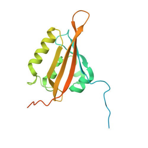
Steric and Electronic Interactions at Gln154 in ZEITLUPE Induce Reorganization of the LOV Domain Dimer Interface.

Pudasaini, A.Green, R.Song, Y.H.Blumenfeld, A.Karki, N.Imaizumi, T.Zoltowski, B.D.

(2021) Biochemistry 60: 95-103

  • DOI: https://doi.org/10.1021/acs.biochem.0c00819
  • Primary Citation of Related Structures:  
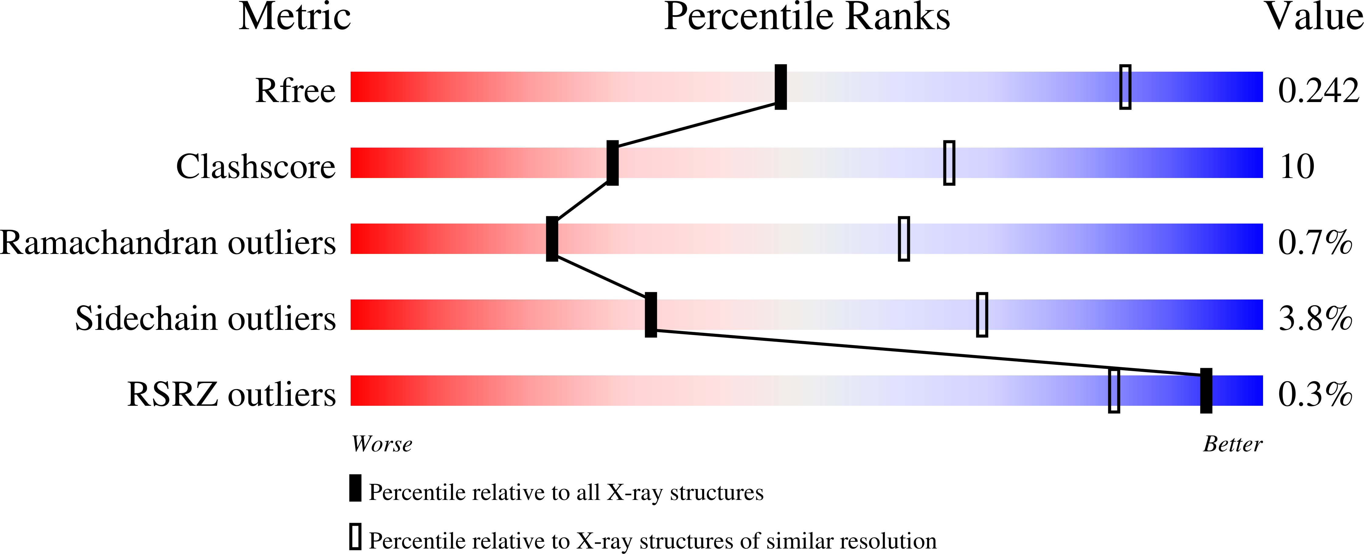
    6WLE, 6WLP

  • PubMed Abstract: 

    Plants measure light quality, intensity, and duration to coordinate growth and development with daily and seasonal changes in environmental conditions; however, the molecular details linking photochemistry to signal transduction remain incomplete. Two closely related light, oxygen, or voltage (LOV) domain-containing photoreceptor proteins, ZEITLUPE (ZTL) and FLAVIN-BINDING, KELCH REPEAT, F-BOX 1 (FKF1), divergently regulate the protein stability of circadian clock and photoperiodic flowering components to mediate daily and seasonal development. Using structural approaches, we identified that mutations at the Gly46 position led to global rearrangements of the ZTL dimer interface in the isolated ZTL-LOV domain. Specifically, G46S and G46A variants induce a 180° rotation about the ZTL-LOV dimer interface that is coupled to ordering of N- and C-terminal signaling elements. These conformational changes hinge upon rotation of a C-terminal Gln residue (Gln154) analogous to that present in light-state structures of ZTL. In contrast to other LOV proteins, a Q154L variant retains light-state interactions with GIGANTEA (GI), thereby indicating N5 protonation is not required for ZTL signaling. The results presented herein confirm a divergent signaling mechanism within ZTL, whereby steric and electronic effects following adduct formation can be sufficient for signal propagation in LOV proteins containing a Gly residue at position 46. Examination of bacterial LOV structures with Gly residues at the equivalent position suggests that mechanisms of signal transduction in LOV proteins may be fluid across the LOV protein family.


  • Organizational Affiliation

    Department of Chemistry, Southern Methodist University, Dallas, Texas 75275, United States.


Macromolecules
Find similar proteins by:  (by identity cutoff)  |  3D Structure
Entity ID: 1
MoleculeChains Sequence LengthOrganismDetailsImage
Adagio protein 1190Arabidopsis thalianaMutation(s): 1 
Gene Names: ADO1FKL2LKP1ZTLAt5g57360MSF19.2
UniProt
Find proteins for Q94BT6 (Arabidopsis thaliana)
Explore Q94BT6 
Go to UniProtKB:  Q94BT6
Entity Groups  
Sequence Clusters30% Identity50% Identity70% Identity90% Identity95% Identity100% Identity
UniProt GroupQ94BT6
Sequence Annotations
Expand
  • Reference Sequence
Small Molecules
Ligands 2 Unique
IDChains Name / Formula / InChI Key2D Diagram3D Interactions
FMN
Query on FMN

Download Ideal Coordinates CCD File 
H [auth B]
I [auth A]
K [auth C]
M [auth D]
O [auth E]
H [auth B],
I [auth A],
K [auth C],
M [auth D],
O [auth E],
P [auth F],
Q [auth G]
FLAVIN MONONUCLEOTIDE
C17 H21 N4 O9 P
FVTCRASFADXXNN-SCRDCRAPSA-N
EDO
Query on EDO

Download Ideal Coordinates CCD File 
J [auth C],
L [auth D],
N [auth E]
1,2-ETHANEDIOL
C2 H6 O2
LYCAIKOWRPUZTN-UHFFFAOYSA-N
Experimental Data & Validation

Experimental Data

  • Method: X-RAY DIFFRACTION
  • Resolution: 3.00 Å
  • R-Value Free: 0.242 
  • R-Value Work: 0.203 
  • R-Value Observed: 0.204 
  • Space Group: I 21 3
Unit Cell:
Length ( Å )Angle ( ˚ )
a = 265.822α = 90
b = 265.822β = 90
c = 265.822γ = 90
Software Package:
Software NamePurpose
PHENIXrefinement
HKL-2000data reduction
HKL-2000data scaling
PHENIXphasing

Structure Validation

View Full Validation Report


Ligand Structure Quality Assessment 


Entry History & Funding Information

Deposition Data


Funding OrganizationLocationGrant Number
National Institutes of Health/National Institute of General Medical Sciences (NIH/NIGMS)United States2R15GM109282

Revision History  (Full details and data files)

  • Version 1.0: 2021-03-03
    Type: Initial release
  • Version 1.1: 2023-10-18
    Changes: Data collection, Database references, Refinement description