6WBH

Crystal structure of mRECK(CC4) in fusion with engineered MBP at medium resolution


Experimental Data Snapshot

  • Method: X-RAY DIFFRACTION
  • Resolution: 2.46 Å
  • R-Value Free: 0.220 
  • R-Value Work: 0.194 
  • R-Value Observed: 0.196 

wwPDB Validation   3D Report Full Report


This is version 2.1 of the entry. See complete history


Literature

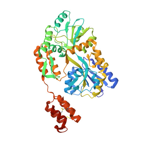
Structure of the RECK CC domain, an evolutionary anomaly.

Chang, T.H.Hsieh, F.L.Smallwood, P.M.Gabelli, S.B.Nathans, J.

(2020) Proc Natl Acad Sci U S A 117: 15104-15111

  • DOI: https://doi.org/10.1073/pnas.2006332117
  • Primary Citation of Related Structures:  
    6WBH, 6WBJ

  • PubMed Abstract: 

    Five small protein domains, the CC-domains, at the N terminus of the RECK protein, play essential roles in signaling by WNT7A and WNT7B in the context of central nervous system angiogenesis and blood-brain barrier formation and maintenance. We have determined the structure of CC domain 4 (CC4) at 1.65-Å resolution and find that it folds into a compact four-helix bundle with three disulfide bonds. The CC4 structure, together with homology modeling of CC1, reveals the surface locations of critical residues that were shown in previous mutagenesis studies to mediate GPR124 binding and WNT7A/WNT7B recognition and signaling. Surprisingly, sequence and structural homology searches reveal no other cell-surface or secreted domains in vertebrates that resemble the CC domain, a pattern that is in striking contrast to other ancient and similarly sized domains, such as Epidermal Growth Factor, Fibronectin Type 3, Immunoglobulin, and Thrombospondin type 1 domains, which are collectively present in hundreds of proteins.


  • Organizational Affiliation

    Department of Molecular Biology and Genetics, Johns Hopkins University School of Medicine, Baltimore, MD 21205.


Macromolecules
Find similar proteins by:  (by identity cutoff)  |  3D Structure
Entity ID: 1
MoleculeChains Sequence LengthOrganismDetailsImage
Maltodextrin-binding protein,Reversion-inducing cysteine-rich protein with Kazal motifs fusion447Escherichia coliMus musculus
This entity is chimeric
Mutation(s): 11 
Gene Names: malEDJ492_13065EPS91_05465FV295_14110NCTC8450_00456Reck
UniProt
Find proteins for Q9Z0J1 (Mus musculus)
Explore Q9Z0J1 
Go to UniProtKB:  Q9Z0J1
Find proteins for P0AEX9 (Escherichia coli (strain K12))
Explore P0AEX9 
Go to UniProtKB:  P0AEX9
Entity Groups  
Sequence Clusters30% Identity50% Identity70% Identity90% Identity95% Identity100% Identity
UniProt GroupsQ9Z0J1P0AEX9
Sequence Annotations
Expand
  • Reference Sequence
Oligosaccharides

Help

Entity ID: 2
MoleculeChains Length2D Diagram Glycosylation3D Interactions
alpha-D-glucopyranose-(1-4)-alpha-D-glucopyranose
B
2N/A
Glycosylation Resources
GlyTouCan:  G07411ON
GlyCosmos:  G07411ON
Biologically Interesting Molecules (External Reference) 1 Unique
Experimental Data & Validation

Experimental Data

  • Method: X-RAY DIFFRACTION
  • Resolution: 2.46 Å
  • R-Value Free: 0.220 
  • R-Value Work: 0.194 
  • R-Value Observed: 0.196 
  • Space Group: P 21 21 2
Unit Cell:
Length ( Å )Angle ( ˚ )
a = 80.222α = 90
b = 110.599β = 90
c = 57.791γ = 90
Software Package:
Software NamePurpose
Aimlessdata scaling
PHASERphasing
PHENIXrefinement
PDB_EXTRACTdata extraction
DIALSdata reduction

Structure Validation

View Full Validation Report


Entry History & Funding Information

Deposition Data


Funding OrganizationLocationGrant Number
National Institutes of Health/National Eye Institute (NIH/NEI)United StatesR01EY018637
Howard Hughes Medical Institute (HHMI)United States--
Human Frontier Science Program (HFSP)FranceLT000130/2017-L

Revision History  (Full details and data files)

  • Version 1.0: 2020-06-17
    Type: Initial release
  • Version 1.1: 2020-07-01
    Changes: Database references
  • Version 1.2: 2020-07-15
    Changes: Database references
  • Version 2.0: 2020-07-29
    Type: Remediation
    Reason: Carbohydrate remediation
    Changes: Atomic model, Author supporting evidence, Data collection, Derived calculations, Non-polymer description, Structure summary
  • Version 2.1: 2023-10-18
    Changes: Author supporting evidence, Data collection, Database references, Refinement description, Structure summary