6WAU

Complex structure of PHF19


Experimental Data Snapshot

  • Method: X-RAY DIFFRACTION
  • Resolution: 1.75 Å
  • R-Value Free: 0.250 
  • R-Value Work: 0.213 

wwPDB Validation   3D Report Full Report


This is version 1.3 of the entry. See complete history


Literature

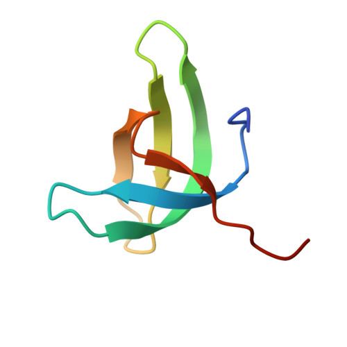
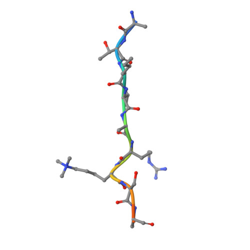
Structural basis for histone variant H3tK27me3 recognition by PHF1 and PHF19.

Dong, C.Nakagawa, R.Oyama, K.Yamamoto, Y.Zhang, W.Dong, A.Li, Y.Yoshimura, Y.Kamiya, H.Nakayama, J.I.Ueda, J.Min, J.

(2020) Elife 9

  • DOI: https://doi.org/10.7554/eLife.58675
  • Primary Citation of Related Structures:  
    6WAT, 6WAU, 6WAV

  • PubMed Abstract: 

    The Polycomb repressive complex 2 (PRC2) is a multicomponent histone H3K27 methyltransferase complex, best known for silencing the Hox genes during embryonic development. The Polycomb-like proteins PHF1, MTF2, and PHF19 are critical components of PRC2 by stimulating its catalytic activity in embryonic stem cells. The Tudor domains of PHF1/19 have been previously shown to be readers of H3K36me3 in vitro. However, some other studies suggest that PHF1 and PHF19 co-localize with the H3K27me3 mark but not H3K36me3 in cells. Here, we provide further evidence that PHF1 co-localizes with H3t in testis and its Tudor domain preferentially binds to H3tK27me3 over canonical H3K27me3 in vitro. Our complex structures of the Tudor domains of PHF1 and PHF19 with H3tK27me3 shed light on the molecular basis for preferential recognition of H3tK27me3 by PHF1 and PHF19 over canonical H3K27me3, implicating that H3tK27me3 might be a physiological ligand of PHF1/19.


  • Organizational Affiliation

    Department of Biochemistry and Molecular Biology, School of Basic Medical Sciences, Tianjin Medical University, Tianjin, China.


Macromolecules
Find similar proteins by:  (by identity cutoff)  |  3D Structure
Entity ID: 1
MoleculeChains Sequence LengthOrganismDetailsImage
PHD finger protein 19
A, B, C, D, E
A, B, C, D, E, F
60Homo sapiensMutation(s): 0 
Gene Names: PHF19PCL3
UniProt & NIH Common Fund Data Resources
Find proteins for Q5T6S3 (Homo sapiens)
Explore Q5T6S3 
Go to UniProtKB:  Q5T6S3
PHAROS:  Q5T6S3
GTEx:  ENSG00000119403 
Entity Groups  
Sequence Clusters30% Identity50% Identity70% Identity90% Identity95% Identity100% Identity
UniProt GroupQ5T6S3
Sequence Annotations
Expand
  • Reference Sequence

Find similar proteins by:  Sequence   |   3D Structure  

Entity ID: 2
MoleculeChains Sequence LengthOrganismDetailsImage
Histone H3.1t peptide
G, H, I, J, K
G, H, I, J, K, L
12Homo sapiensMutation(s): 0 
UniProt & NIH Common Fund Data Resources
Find proteins for Q16695 (Homo sapiens)
Explore Q16695 
Go to UniProtKB:  Q16695
PHAROS:  Q16695
GTEx:  ENSG00000168148 
Entity Groups  
Sequence Clusters30% Identity50% Identity70% Identity90% Identity95% Identity100% Identity
UniProt GroupQ16695
Sequence Annotations
Expand
  • Reference Sequence
Small Molecules
Ligands 1 Unique
IDChains Name / Formula / InChI Key2D Diagram3D Interactions
UNX
Query on UNX

Download Ideal Coordinates CCD File 
M [auth A]
N [auth B]
O [auth B]
P [auth B]
Q [auth B]
M [auth A],
N [auth B],
O [auth B],
P [auth B],
Q [auth B],
R [auth C],
S [auth C],
T [auth D],
U [auth D],
V [auth D],
W [auth D]
UNKNOWN ATOM OR ION
X
Modified Residues  1 Unique
IDChains TypeFormula2D DiagramParent
M3L
Query on M3L
G, H, I, J, K
G, H, I, J, K, L
L-PEPTIDE LINKINGC9 H21 N2 O2LYS
Experimental Data & Validation

Experimental Data

Unit Cell:
Length ( Å )Angle ( ˚ )
a = 111.477α = 90
b = 111.477β = 90
c = 34.409γ = 120
Software Package:
Software NamePurpose
Aimlessdata scaling
REFMACrefinement
PDB_EXTRACTdata extraction
XDSdata reduction
PHASERphasing

Structure Validation

View Full Validation Report


Entry History 

Deposition Data

Revision History  (Full details and data files)

  • Version 1.0: 2020-08-26
    Type: Initial release
  • Version 1.1: 2020-09-16
    Changes: Database references
  • Version 1.2: 2021-06-30
    Changes: Data collection
  • Version 1.3: 2023-10-18
    Changes: Data collection, Database references, Refinement description