6WA6

Structure of the Chlamydia pneumoniae CdsV protein


Experimental Data Snapshot

  • Method: X-RAY DIFFRACTION
  • Resolution: 2.80 Å
  • R-Value Free: 0.244 
  • R-Value Work: 0.196 
  • R-Value Observed: 0.198 

wwPDB Validation   3D Report Full Report


This is version 1.2 of the entry. See complete history


Literature

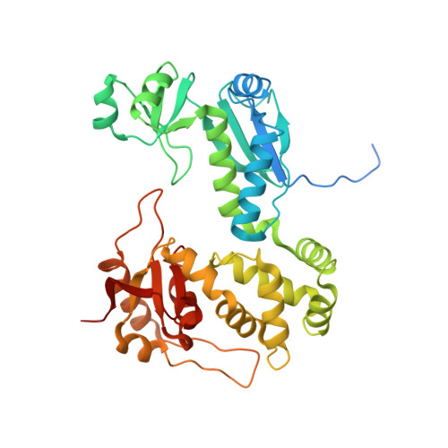
"The structure of the Type III secretion system export gate with CdsO, an ATPase lever arm".

Jensen, J.L.Yamini, S.Rietsch, A.Spiller, B.W.

(2020) PLoS Pathog 16: e1008923-e1008923

  • DOI: https://doi.org/10.1371/journal.ppat.1008923
  • Primary Citation of Related Structures:  
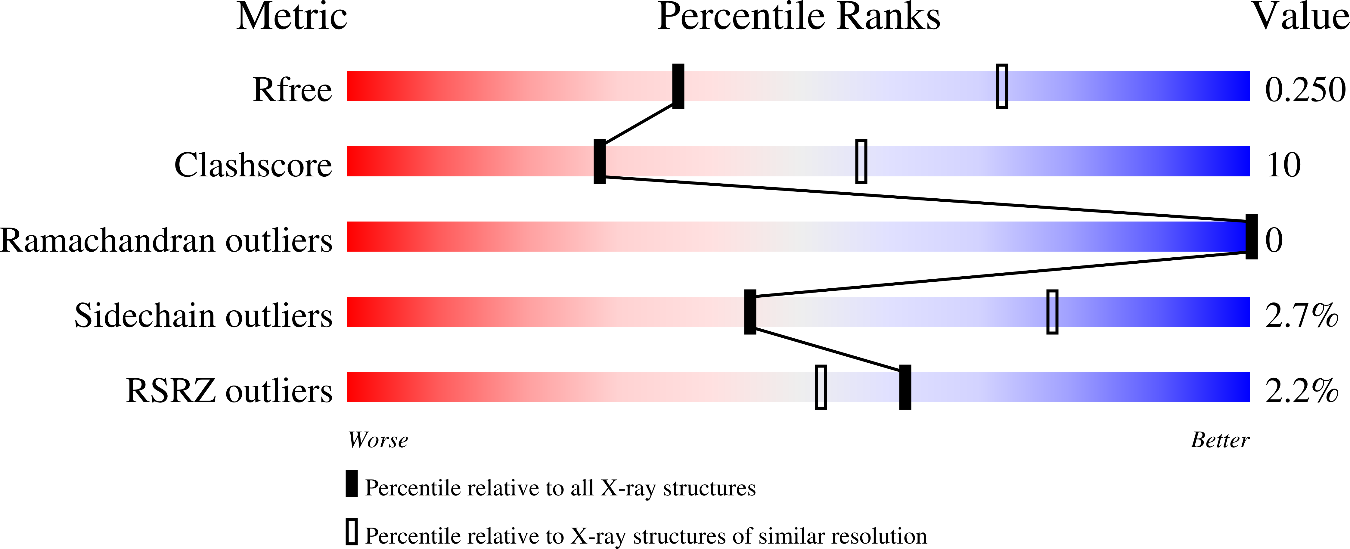
    6WA6, 6WA9

  • PubMed Abstract: 

    Type III protein secretion systems (T3SS) deliver effector proteins from the Gram-negative bacterial cytoplasm into a eukaryotic host cell through a syringe-like, multi-protein nanomachine. Cytosolic components of T3SS include a portion of the export apparatus, which traverses the inner membrane and features the opening of the secretion channel, and the sorting complex for substrate recognition and for providing the energetics required for protein secretion. Two components critical for efficient effector export are the export gate protein and the ATPase, which are proposed to be linked by the central stalk protein of the ATPase. We present the structure of the soluble export gate homo-nonamer, CdsV, in complex with the central stalk protein, CdsO, of its cognate ATPase, both derived from Chlamydia pneumoniae. This structure defines the interface between these essential T3S proteins and reveals that CdsO engages the periphery of the export gate that may allow the ATPase to catalyze an opening between export gate subunits to allow cargo to enter the export apparatus. We also demonstrate through structure-based mutagenesis of the homologous export gate in Pseudomonas aeruginosa that mutation of this interface disrupts effector secretion. These results provide novel insights into the molecular mechanisms governing active substrate recognition and translocation through a T3SS.


  • Organizational Affiliation

    Department of Pathology, Microbiology and Immunology, Vanderbilt University Medical Center, Nashville, TN, United States of America.


Macromolecules
Find similar proteins by:  (by identity cutoff)  |  3D Structure
Entity ID: 1
MoleculeChains Sequence LengthOrganismDetailsImage
Low calcium response locus protein D
A, B, C, D, E
A, B, C, D, E, F, G, H, I
387Chlamydia pneumoniaeMutation(s): 0 
Gene Names: 
UniProt
Find proteins for Q9Z8L5 (Chlamydia pneumoniae)
Explore Q9Z8L5 
Go to UniProtKB:  Q9Z8L5
Entity Groups  
Sequence Clusters30% Identity50% Identity70% Identity90% Identity95% Identity100% Identity
UniProt GroupQ9Z8L5
Sequence Annotations
Expand
  • Reference Sequence
Small Molecules
Ligands 1 Unique
IDChains Name / Formula / InChI Key2D Diagram3D Interactions
GOL
Query on GOL

Download Ideal Coordinates CCD File 
J [auth A]
K [auth A]
L [auth A]
M [auth A]
N [auth B]
J [auth A],
K [auth A],
L [auth A],
M [auth A],
N [auth B],
O [auth B],
P [auth C],
Q [auth D],
R [auth E],
S [auth E],
T [auth E],
U [auth G],
V [auth I]
GLYCEROL
C3 H8 O3
PEDCQBHIVMGVHV-UHFFFAOYSA-N
Experimental Data & Validation

Experimental Data

  • Method: X-RAY DIFFRACTION
  • Resolution: 2.80 Å
  • R-Value Free: 0.244 
  • R-Value Work: 0.196 
  • R-Value Observed: 0.198 
  • Space Group: P 21 21 21
Unit Cell:
Length ( Å )Angle ( ˚ )
a = 66.65α = 90
b = 280.44β = 90
c = 290.41γ = 90
Software Package:
Software NamePurpose
PHENIXrefinement
XDSdata reduction
XDSdata scaling
SHARPphasing

Structure Validation

View Full Validation Report


Entry History & Funding Information

Deposition Data


Funding OrganizationLocationGrant Number
National Institutes of Health/National Institute Of Allergy and Infectious Diseases (NIH/NIAID)United States1R01AI108778

Revision History  (Full details and data files)

  • Version 1.0: 2020-09-30
    Type: Initial release
  • Version 1.1: 2020-10-28
    Changes: Database references
  • Version 1.2: 2024-03-06
    Changes: Data collection, Database references