6VUG

Diabody bound to a Reverse Transcriptase Aptamer Complex


Experimental Data Snapshot

  • Method: X-RAY DIFFRACTION
  • Resolution: 3.00 Å
  • R-Value Free: 0.255 
  • R-Value Work: 0.202 
  • R-Value Observed: 0.205 

wwPDB Validation   3D Report Full Report


This is version 1.3 of the entry. See complete history


Literature

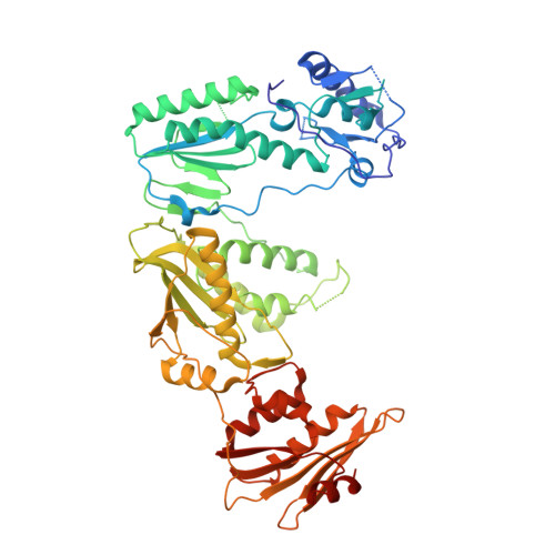
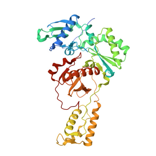
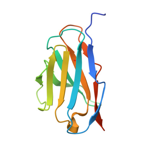
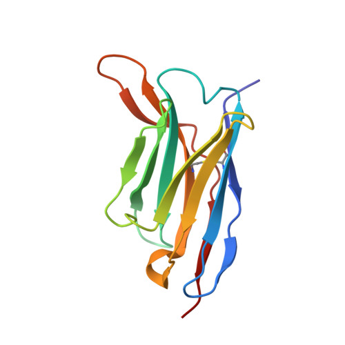
Co-crystallization with diabodies: A case study for the introduction of synthetic symmetry.

Chesterman, C.Arnold, E.

(2021) Structure 29: 598-605.e3

  • DOI: https://doi.org/10.1016/j.str.2021.02.001
  • Primary Citation of Related Structures:  
    6VRP, 6VUG, 6VUN, 6VUO, 6VUP, 7KBM, 7KBO, 7KBP

  • PubMed Abstract: 

    This work presents a method for introducing synthetic symmetry into protein crystallization samples using an antibody fragment termed a diabody (Dab). These Dabs contain two target binding sites, and engineered disulfide bonds have been included to modulate Dab flexibility. The impacts of Dab engineering have been observed through assessment of thermal stability, small-angle X-ray scattering, and high-resolution crystal structures. Complexes between the engineered Dabs and HIV-1 reverse transcriptase (RT) bound to a high-affinity DNA aptamer were also generated to explore the capacity of engineered Dabs to enable the crystallization of bound target proteins. This strategy increased the crystallization hit frequency obtained for RT-aptamer, and the structure of a Dab-RT-aptamer complex was determined to 3.0-Å resolution. Introduction of synthetic symmetry using a Dab could be a broadly applicable strategy, especially when monoclonal antibodies for a target have previously been identified.


  • Organizational Affiliation

    Center for Advanced Biotechnology and Medicine and Department of Chemistry and Chemical Biology, Rutgers University, Piscataway, NJ 08854, USA; GSK, Rockville, MD 20850, USA.


Macromolecules

Find similar proteins by:  (by identity cutoff)  |  3D Structure
Entity ID: 1
MoleculeChains Sequence LengthOrganismDetailsImage
Reverse transcriptase/ribonuclease H555Human immunodeficiency virus 1Mutation(s): 0 
Gene Names: gag-pol
EC: 3.4.23.16 (PDB Primary Data), 2.7.7.49 (PDB Primary Data), 2.7.7.7 (PDB Primary Data), 3.1.26.13 (PDB Primary Data), 3.1.13.2 (PDB Primary Data), 2.7.7 (PDB Primary Data), 3.1 (PDB Primary Data)
UniProt
Find proteins for P03366 (Human immunodeficiency virus type 1 group M subtype B (isolate BH10))
Explore P03366 
Go to UniProtKB:  P03366
Entity Groups  
Sequence Clusters30% Identity50% Identity70% Identity90% Identity95% Identity100% Identity
UniProt GroupP03366
Sequence Annotations
Expand
  • Reference Sequence
Find similar proteins by:  (by identity cutoff)  |  3D Structure
Entity ID: 2
MoleculeChains Sequence LengthOrganismDetailsImage
REVERSE TRANSCRIPTASE P51444Human immunodeficiency virus 1Mutation(s): 2 
Gene Names: pol
EC: 2.7.7.49 (PDB Primary Data), 3.1.13.2 (PDB Primary Data), 3.1.26.13 (PDB Primary Data)
UniProt
Find proteins for P03366 (Human immunodeficiency virus type 1 group M subtype B (isolate BH10))
Explore P03366 
Go to UniProtKB:  P03366
Entity Groups  
Sequence Clusters30% Identity50% Identity70% Identity90% Identity95% Identity100% Identity
UniProt GroupP03366
Sequence Annotations
Expand
  • Reference Sequence
Find similar proteins by:  (by identity cutoff)  |  3D Structure
Entity ID: 3
MoleculeChains Sequence LengthOrganismDetailsImage
Light chain variable fragment120Homo sapiensMutation(s): 0 
Entity Groups  
Sequence Clusters30% Identity50% Identity70% Identity90% Identity95% Identity100% Identity
Sequence Annotations
Expand
  • Reference Sequence
Find similar proteins by:  (by identity cutoff)  |  3D Structure
Entity ID: 4
MoleculeChains Sequence LengthOrganismDetailsImage
Heavy chain variable fragment124Homo sapiensMutation(s): 0 
Entity Groups  
Sequence Clusters30% Identity50% Identity70% Identity90% Identity95% Identity100% Identity
Sequence Annotations
Expand
  • Reference Sequence
Find similar nucleic acids by:  (by identity cutoff)  |  3D Structure
Entity ID: 5
MoleculeChains LengthOrganismImage
DNA (38-MER)38Human immunodeficiency virus 1
Sequence Annotations
Expand
  • Reference Sequence
Small Molecules
Ligands 2 Unique
IDChains Name / Formula / InChI Key2D Diagram3D Interactions
SO4
Query on SO4

Download Ideal Coordinates CCD File 
F [auth B]
G [auth B]
H [auth B]
I [auth B]
J [auth B]
F [auth B],
G [auth B],
H [auth B],
I [auth B],
J [auth B],
K [auth B],
L [auth B],
M [auth B],
O [auth C],
P [auth C],
Q [auth C]
SULFATE ION
O4 S
QAOWNCQODCNURD-UHFFFAOYSA-L
GOL
Query on GOL

Download Ideal Coordinates CCD File 
N [auth B]GLYCEROL
C3 H8 O3
PEDCQBHIVMGVHV-UHFFFAOYSA-N
Experimental Data & Validation

Experimental Data

  • Method: X-RAY DIFFRACTION
  • Resolution: 3.00 Å
  • R-Value Free: 0.255 
  • R-Value Work: 0.202 
  • R-Value Observed: 0.205 
  • Space Group: C 2 2 21
Unit Cell:
Length ( Å )Angle ( ˚ )
a = 85.876α = 90
b = 238.103β = 90
c = 211.711γ = 90
Software Package:
Software NamePurpose
PHENIXrefinement
HKL-2000data reduction
HKL-2000data scaling
PHASERphasing

Structure Validation

View Full Validation Report


Entry History & Funding Information

Deposition Data


Funding OrganizationLocationGrant Number
National Institutes of Health/National Institute of General Medical Sciences (NIH/NIGMS)United StatesR37 AI027690

Revision History  (Full details and data files)

  • Version 1.0: 2021-02-17
    Type: Initial release
  • Version 1.1: 2021-03-10
    Changes: Database references
  • Version 1.2: 2021-06-16
    Changes: Database references
  • Version 1.3: 2023-10-11
    Changes: Data collection, Database references, Refinement description