6VP3

Artificial Metalloproteins with Dinuclear Iron Centers


Experimental Data Snapshot

  • Method: X-RAY DIFFRACTION
  • Resolution: 1.65 Å
  • R-Value Free: 0.221 
  • R-Value Work: 0.176 
  • R-Value Observed: 0.178 

wwPDB Validation   3D Report Full Report


Ligand Structure Quality Assessment 


This is version 1.3 of the entry. See complete history


Literature

Artificial Metalloproteins with Dinuclear Iron-Hydroxido Centers.

Miller, K.R.Biswas, S.Jasniewski, A.Follmer, A.H.Biswas, A.Albert, T.Sabuncu, S.Bominaar, E.L.Hendrich, M.P.Moenne-Loccoz, P.Borovik, A.S.

(2021) J Am Chem Soc 143: 2384-2393

  • DOI: https://doi.org/10.1021/jacs.0c12564
  • Primary Citation of Related Structures:  
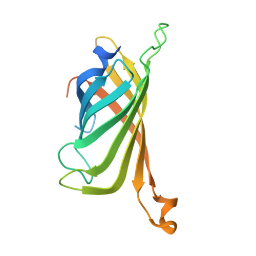
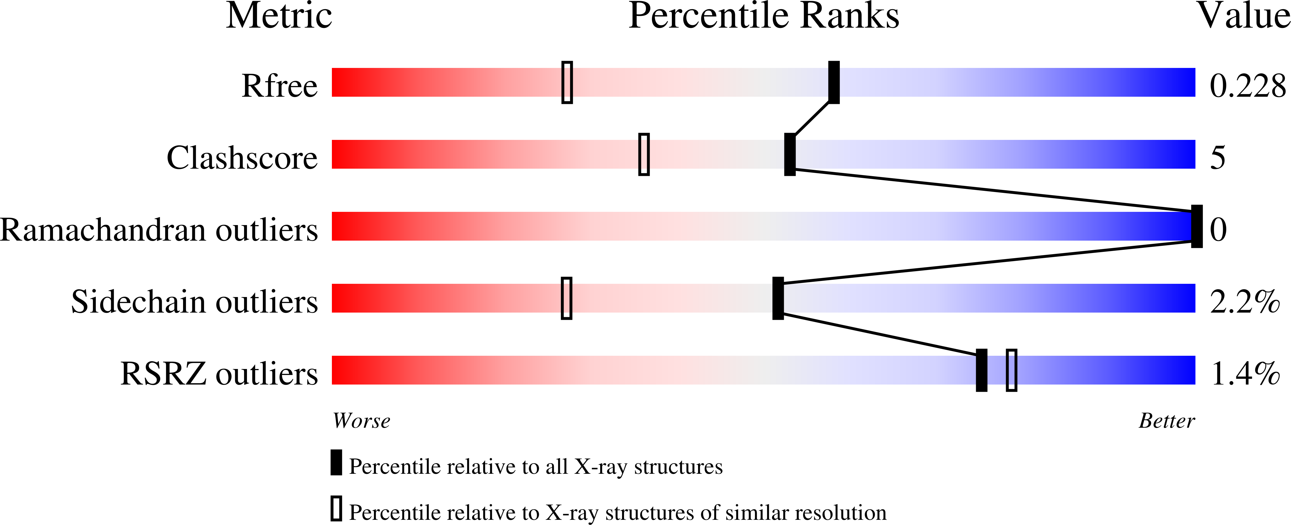
    6VO9, 6VOB, 6VOZ, 6VP1, 6VP2, 6VP3, 7KBY, 7KBZ, 7KNL

  • PubMed Abstract: 

    Dinuclear iron centers with a bridging hydroxido or oxido ligand form active sites within a variety of metalloproteins. A key feature of these sites is the ability of the protein to control the structures around the Fe centers, which leads to entatic states that are essential for function. To simulate this controlled environment, artificial proteins have been engineered using biotin-streptavidin (Sav) technology in which Fe complexes from adjacent subunits can assemble to form [Fe III -(μ-OH)-Fe III ] cores. The assembly process is promoted by the site-specific localization of the Fe complexes within a subunit through the designed mutation of a tyrosinate side chain to coordinate the Fe centers. An important outcome is that the Sav host can regulate the Fe···Fe separation, which is known to be important for function in natural metalloproteins. Spectroscopic and structural studies from X-ray diffraction methods revealed uncommonly long Fe···Fe separations that change by less than 0.3 Å upon the binding of additional bridging ligands. The structural constraints imposed by the protein host on the di-Fe cores are unique and create examples of active sites having entatic states within engineered artificial metalloproteins.


  • Organizational Affiliation

    Department of Chemistry, 1102 Natural Sciences II, University of California, Irvine, California 92697, United States.


Macromolecules
Find similar proteins by:  (by identity cutoff)  |  3D Structure
Entity ID: 1
MoleculeChains Sequence LengthOrganismDetailsImage
Streptavidin
A, B, C, D
159Streptomyces avidiniiMutation(s): 2 
UniProt
Find proteins for P22629 (Streptomyces avidinii)
Explore P22629 
Go to UniProtKB:  P22629
Entity Groups  
Sequence Clusters30% Identity50% Identity70% Identity90% Identity95% Identity100% Identity
UniProt GroupP22629
Sequence Annotations
Expand
  • Reference Sequence
Experimental Data & Validation

Experimental Data

  • Method: X-RAY DIFFRACTION
  • Resolution: 1.65 Å
  • R-Value Free: 0.221 
  • R-Value Work: 0.176 
  • R-Value Observed: 0.178 
  • Space Group: C 1 2 1
Unit Cell:
Length ( Å )Angle ( ˚ )
a = 193.503α = 90
b = 57.861β = 107.4
c = 57.861γ = 90
Software Package:
Software NamePurpose
Aimlessdata scaling
REFMACrefinement
PDB_EXTRACTdata extraction
XDSdata reduction
PHASERphasing

Structure Validation

View Full Validation Report


Ligand Structure Quality Assessment 


Entry History & Funding Information

Deposition Data


Funding OrganizationLocationGrant Number
National Institutes of Health/National Institute of General Medical Sciences (NIH/NIGMS)United StatesGM120349

Revision History  (Full details and data files)

  • Version 1.0: 2021-02-03
    Type: Initial release
  • Version 1.1: 2021-02-17
    Changes: Database references
  • Version 1.2: 2021-02-24
    Changes: Database references
  • Version 1.3: 2023-10-11
    Changes: Data collection, Database references, Refinement description