6VNU

X-ray Crystal Structure of Ruthenocenyl-7-Aminocephalosporanic Acid Covalent Acyl-Enzyme Complex with CTX-M-14 E166A Beta-Lactamase


Experimental Data Snapshot

  • Method: X-RAY DIFFRACTION
  • Resolution: 1.47 Å
  • R-Value Free: 0.218 
  • R-Value Work: 0.173 
  • R-Value Observed: 0.175 

wwPDB Validation   3D Report Full Report

Currently 6VNU does not have a validation slider image.


This is version 1.2 of the entry. See complete history


Literature

Metallocenyl 7-ACA Conjugates: Antibacterial Activity Studies and Atomic-Resolution X-ray Crystal Structure with CTX-M beta-Lactamase.

Lewandowski, E.M.Szczupak, L.Kowalczyk, A.Mendoza, G.Arruebo, M.Jacobs, L.M.C.Staczek, P.Chen, Y.Kowalski, K.

(2020) Chembiochem 21: 2187-2195

  • DOI: https://doi.org/10.1002/cbic.202000054
  • Primary Citation of Related Structures:  
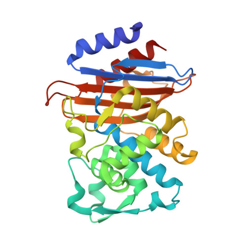
    6VNU

  • PubMed Abstract: 

    The conjugation of organometallic groups to current β-lactam antibiotics is a field of increasing study due to the ability of certain organometallic groups to enhance the antibiotic potency of these drugs. Herein, we report the antibacterial properties of two metallocenyl (ferrocenyl and ruthenocenyl) 7-aminocephalosporanic acid (7-ACA) antibiotic conjugates. Continuing a trend we found in our previous studies, the ruthenocenyl conjugate showed greater antibacterial activity than its ferrocenyl counterpart. Compared with the previously published 7-aminodesacetoxycephalosporanic acid (7-ADCA) conjugates, the 3-acetyloxymethyl group significantly improved the compounds' activity. Furthermore, the Rc-7-ACA compound was more active against clinical Staphylococcus aureus isolates than the ampicillin reference. Noticeably, neither of the two new compounds showed an undesirable toxic effect in HeLa and L929 cells at the concentrations at which they displayed strong antibacterial effects. The antibacterial activity of the two metallocenyl 7-ACA derivatives was further confirmed by scanning electron microscopy (SEM). SEM micrographs showed that bacteria treated with metallocenyl 7-ACA derivatives feature cell wall damage and morphology changes. Using a CTX-M-14 β-lactamase competition assay based on nitrocefin hydrolysis, we showed that the Rc-7-ACA bound more favorably to CTX-M-14 than its ferrocenyl counterpart, again confirming the superiority of the ruthenocenyl moiety over the ferrocenyl one in interacting with proteins. We also report a 1.47 Å resolution crystal structure of Rc-7-ACA in complex with the CTX-M-14 E166A mutant, an enzyme sharing a similar active site configuration with penicillin-binding proteins, the molecular target of β-lactam antibiotics. These results strengthen the case for the antibacterial utility of the Rc and Fc groups.


  • Organizational Affiliation

    Department of Molecular Medicine, University of South Florida, >Morsani College of Medicine, 12901 Bruce B. Downs Boulevard, Tampa, FL, 33612, US.


Macromolecules
Find similar proteins by:  (by identity cutoff)  |  3D Structure
Entity ID: 1
MoleculeChains Sequence LengthOrganismDetailsImage
Beta-lactamase
A, B
263Escherichia coliMutation(s): 1 
Gene Names: blaCTX-MCTX-M-14
EC: 3.5.2.6
UniProt
Find proteins for Q9L5C7 (Escherichia coli)
Explore Q9L5C7 
Go to UniProtKB:  Q9L5C7
Entity Groups  
Sequence Clusters30% Identity50% Identity70% Identity90% Identity95% Identity100% Identity
UniProt GroupQ9L5C7
Sequence Annotations
Expand
  • Reference Sequence
Small Molecules
Ligands 3 Unique
IDChains Name / Formula / InChI Key2D Diagram3D Interactions
TDJ (Subject of Investigation/LOI)
Query on TDJ

Download Ideal Coordinates CCD File 
E [auth A],
H [auth B]
[(1,2,3,4,5-eta)-1-(4-{[carboxy(4-carboxy-5-methylidene-5,6-dihydro-2H-1,3-thiazin-2-yl)methyl]amino}-4-oxobutanoyl)cyclopentadienyl][(1,2,3,4,5-eta)-cyclopentadienyl]ruthenium
C22 H13 N2 O6 Ru S
NNOXJZPSFXUGDA-FZMMWMHASA-N
7G4 (Subject of Investigation/LOI)
Query on 7G4

Download Ideal Coordinates CCD File 
C [auth A],
D [auth A],
G [auth B]
ruthenocene
C10 H10 Ru
BKEJVRMLCVMJLG-UHFFFAOYSA-N
K
Query on K

Download Ideal Coordinates CCD File 
F [auth A],
I [auth B]
POTASSIUM ION
K
NPYPAHLBTDXSSS-UHFFFAOYSA-N
Experimental Data & Validation

Experimental Data

  • Method: X-RAY DIFFRACTION
  • Resolution: 1.47 Å
  • R-Value Free: 0.218 
  • R-Value Work: 0.173 
  • R-Value Observed: 0.175 
  • Space Group: P 1 21 1
Unit Cell:
Length ( Å )Angle ( ˚ )
a = 45.152α = 90
b = 106.894β = 101.68
c = 47.908γ = 90
Software Package:
Software NamePurpose
REFMACrefinement
PDB_EXTRACTdata extraction
HKL-2000data reduction
SCALEPACKdata scaling
PHASERphasing

Structure Validation

View Full Validation Report

Currently 6VNU does not have a validation slider image.


Entry History & Funding Information

Deposition Data


Funding OrganizationLocationGrant Number
National Institutes of Health/National Institute Of Allergy and Infectious Diseases (NIH/NIAID)United StatesAI147654
Polish National Science CentrePolandOPUS UMO-2018/29/B/ST5/00055

Revision History  (Full details and data files)

  • Version 1.0: 2020-04-01
    Type: Initial release
  • Version 1.1: 2020-08-12
    Changes: Database references, Derived calculations
  • Version 1.2: 2023-10-11
    Changes: Data collection, Database references, Refinement description