6V8X

VRC01 Bound BG505 F14 HIV-1 SOSIP Envelope Trimer Structure


Experimental Data Snapshot

  • Method: ELECTRON MICROSCOPY
  • Resolution: 3.00 Å
  • Aggregation State: PARTICLE 
  • Reconstruction Method: SINGLE PARTICLE 

wwPDB Validation   3D Report Full Report


This is version 2.0 of the entry. See complete history


Literature

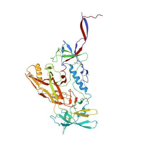
Disruption of the HIV-1 Envelope allosteric network blocks CD4-induced rearrangements.

Henderson, R.Lu, M.Zhou, Y.Mu, Z.Parks, R.Han, Q.Hsu, A.L.Carter, E.Blanchard, S.C.Edwards, R.J.Wiehe, K.Saunders, K.O.Borgnia, M.J.Bartesaghi, A.Mothes, W.Haynes, B.F.Acharya, P.Munir Alam, S.

(2020) Nat Commun 11: 520-520

  • DOI: https://doi.org/10.1038/s41467-019-14196-w
  • Primary Citation of Related Structures:  
    6V8X, 6V8Z

  • PubMed Abstract: 

    The trimeric HIV-1 Envelope protein (Env) mediates viral-host cell fusion via a network of conformational transitions, with allosteric elements in each protomer orchestrating host receptor-induced exposure of the co-receptor binding site and fusion elements. To understand the molecular details of this allostery, here, we introduce Env mutations aimed to prevent CD4-induced rearrangements in the HIV-1 BG505 Env trimer. Binding analysis and single-molecule Förster Resonance Energy Transfer confirm that these mutations prevent CD4-induced transitions of the HIV-1 Env. Structural analysis by single-particle cryo-electron microscopy performed on the BG505 SOSIP mutant Env proteins shows rearrangements in the gp120 topological layer contacts with gp41. Displacement of a conserved tryptophan (W571) from its typical pocket in these Env mutants renders the Env insensitive to CD4 binding. These results reveal the critical function of W571 as a conformational switch in Env allostery and receptor-mediated viral entry and provide insights on Env conformation that are relevant for vaccine design.


  • Organizational Affiliation

    Department of Medicine, Duke University School of Medicine, Durham, NC, 27710, USA. rory.henderson@duke.edu.


Macromolecules
Find similar proteins by:  (by identity cutoff)  |  3D Structure
Entity ID: 1
MoleculeChains Sequence LengthOrganismDetailsImage
Envelope glycoprotein gp120
A, E, I
471Human immunodeficiency virus 1Mutation(s): 0 
Gene Names: env
UniProt
Find proteins for Q2N0S6 (Human immunodeficiency virus 1)
Explore Q2N0S6 
Go to UniProtKB:  Q2N0S6
Entity Groups  
Sequence Clusters30% Identity50% Identity70% Identity90% Identity95% Identity100% Identity
UniProt GroupQ2N0S6
Sequence Annotations
Expand
  • Reference Sequence
Find similar proteins by:  (by identity cutoff)  |  3D Structure
Entity ID: 2
MoleculeChains Sequence LengthOrganismDetailsImage
Envelope glycoprotein gp41
B, F, J
145Human immunodeficiency virus 1Mutation(s): 0 
Gene Names: env
UniProt
Find proteins for Q2N0S9 (Human immunodeficiency virus 1)
Explore Q2N0S9 
Go to UniProtKB:  Q2N0S9
Entity Groups  
Sequence Clusters30% Identity50% Identity70% Identity90% Identity95% Identity100% Identity
UniProt GroupQ2N0S9
Sequence Annotations
Expand
  • Reference Sequence
Find similar proteins by:  (by identity cutoff)  |  3D Structure
Entity ID: 3
MoleculeChains Sequence LengthOrganismDetailsImage
VRC01 Fab Heavy Chain
C, G, K
224Homo sapiensMutation(s): 0 
UniProt
Find proteins for Q6N095 (Homo sapiens)
Explore Q6N095 
Go to UniProtKB:  Q6N095
Entity Groups  
Sequence Clusters30% Identity50% Identity70% Identity90% Identity95% Identity100% Identity
UniProt GroupQ6N095
Sequence Annotations
Expand
  • Reference Sequence
Find similar proteins by:  (by identity cutoff)  |  3D Structure
Entity ID: 4
MoleculeChains Sequence LengthOrganismDetailsImage
VRC01 Fab Light Chain
D, H, L
208Homo sapiensMutation(s): 0 
UniProt
Find proteins for Q6PIL8 (Homo sapiens)
Explore Q6PIL8 
Go to UniProtKB:  Q6PIL8
Entity Groups  
Sequence Clusters30% Identity50% Identity70% Identity90% Identity95% Identity100% Identity
UniProt GroupQ6PIL8
Sequence Annotations
Expand
  • Reference Sequence
Oligosaccharides

Help

Entity ID: 5
MoleculeChains Length2D Diagram Glycosylation3D Interactions
alpha-D-mannopyranose-(1-3)-beta-D-mannopyranose-(1-4)-2-acetamido-2-deoxy-beta-D-glucopyranose-(1-4)-2-acetamido-2-deoxy-beta-D-glucopyranoseKA [auth k],
M,
Y
4N-Glycosylation
Glycosylation Resources
GlyTouCan:  G81315DD
GlyCosmos:  G81315DD
GlyGen:  G81315DD
Entity ID: 7
MoleculeChains Length2D Diagram Glycosylation3D Interactions
beta-D-mannopyranose-(1-4)-2-acetamido-2-deoxy-beta-D-glucopyranose-(1-4)-2-acetamido-2-deoxy-beta-D-glucopyranose
AA [auth a],
BA [auth b],
CA [auth c],
FA [auth f],
MA [auth m],
AA [auth a],
BA [auth b],
CA [auth c],
FA [auth f],
MA [auth m],
NA [auth n],
O,
OA [auth o],
P,
Q,
RA [auth r],
T
3N-Glycosylation
Glycosylation Resources
GlyTouCan:  G15407YE
GlyCosmos:  G15407YE
GlyGen:  G15407YE
Small Molecules
Ligands 1 Unique
IDChains Name / Formula / InChI Key2D Diagram3D Interactions
NAG
Query on NAG

Download Ideal Coordinates CCD File 
AB [auth B]
BB [auth E]
CB [auth E]
DB [auth E]
EB [auth E]
AB [auth B],
BB [auth E],
CB [auth E],
DB [auth E],
EB [auth E],
FB [auth F],
GB [auth I],
HB [auth I],
IB [auth I],
JB [auth I],
KB [auth J],
WA [auth A],
XA [auth A],
YA [auth A],
ZA [auth A]
2-acetamido-2-deoxy-beta-D-glucopyranose
C8 H15 N O6
OVRNDRQMDRJTHS-FMDGEEDCSA-N
Experimental Data & Validation

Experimental Data

  • Method: ELECTRON MICROSCOPY
  • Resolution: 3.00 Å
  • Aggregation State: PARTICLE 
  • Reconstruction Method: SINGLE PARTICLE 

Structure Validation

View Full Validation Report


Entry History & Funding Information

Deposition Data


Funding OrganizationLocationGrant Number
National Institutes of Health/National Institute Of Allergy and Infectious Diseases (NIH/NIAID)United States1R01AI145687-01

Revision History  (Full details and data files)

  • Version 1.0: 2020-02-05
    Type: Initial release
  • Version 1.1: 2020-02-12
    Changes: Database references
  • Version 2.0: 2020-07-29
    Type: Remediation
    Reason: Carbohydrate remediation
    Changes: Advisory, Atomic model, Data collection, Derived calculations, Structure summary