6V64

Crystal structure of human thrombin bound to ppack with tryptophans replaced by 5-F-tryptophan


Experimental Data Snapshot

  • Method: X-RAY DIFFRACTION
  • Resolution: 2.29 Å
  • R-Value Free: 0.277 
  • R-Value Work: 0.197 
  • R-Value Observed: 0.200 

wwPDB Validation   3D Report Full Report


Ligand Structure Quality Assessment 


This is version 1.2 of the entry. See complete history


Literature

19F NMR reveals the conformational properties of free thrombin and its zymogen precursor prethrombin-2.

Ruben, E.A.Gandhi, P.S.Chen, Z.Koester, S.K.DeKoster, G.T.Frieden, C.Di Cera, E.

(2020) J Biol Chem 295: 8227-8235

  • DOI: https://doi.org/10.1074/jbc.RA120.013419
  • Primary Citation of Related Structures:  
    6V5T, 6V64

  • PubMed Abstract: 

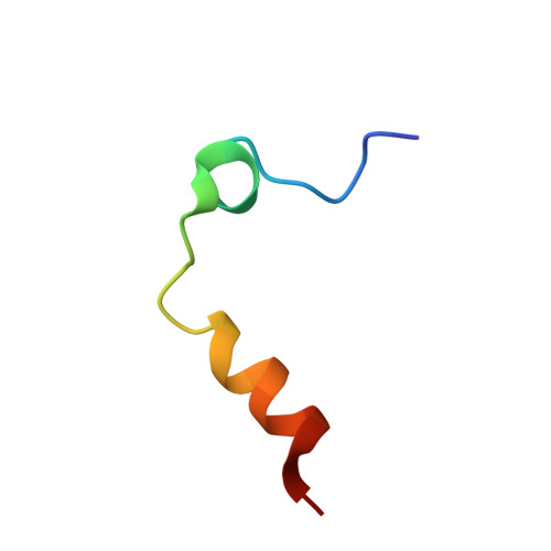
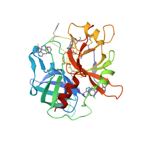
    The conformational properties of trypsin-like proteases and their zymogen forms remain controversial because of a lack of sufficient information on their free forms. Specifically, it is unclear whether the free protease is zymogen-like and shifts to its mature form upon a ligand-induced fit or exists in multiple conformations in equilibrium from which the ligand selects the optimal fit via conformational selection. Here we report the results of 19 F NMR measurements that reveal the conformational properties of a protease and its zymogen precursor in the free form. Using the trypsin-like, clotting protease thrombin as a relevant model system, we show that its conformation is quite different from that of its direct zymogen precursor prethrombin-2 and more similar to that of its fully active Na + -bound form. The results cast doubts on recent hypotheses that free thrombin is zymogen-like and transitions to protease-like forms upon ligand binding. Rather, they validate the scenario emerged from previous findings of X-ray crystallography and rapid kinetics supporting a pre-existing equilibrium between open (E) and closed (E*) forms of the active site. In this scenario, prethrombin-2 is more dynamic and exists predominantly in the E* form, whereas thrombin is more rigid and exists predominantly in the E form. Ligand binding to thrombin takes place exclusively in the E form without significant changes in the overall conformation. In summary, these results disclose the structural architecture of the free forms of thrombin and prethrombin-2, consistent with an E*-E equilibrium and providing no evidence that free thrombin is zymogen-like.


  • Organizational Affiliation

    Edward A. Doisy Department of Biochemistry and Molecular Biology, Saint Louis University School of Medicine, St. Louis, Missouri, USA.


Macromolecules
Find similar proteins by:  (by identity cutoff)  |  3D Structure
Entity ID: 1
MoleculeChains Sequence LengthOrganismDetailsImage
Thrombin light chain30Homo sapiensMutation(s): 0 
Gene Names: F2
EC: 3.4.21.5
UniProt & NIH Common Fund Data Resources
Find proteins for P00734 (Homo sapiens)
Explore P00734 
Go to UniProtKB:  P00734
PHAROS:  P00734
GTEx:  ENSG00000180210 
Entity Groups  
Sequence Clusters30% Identity50% Identity70% Identity90% Identity95% Identity100% Identity
UniProt GroupP00734
Sequence Annotations
Expand
  • Reference Sequence
Find similar proteins by:  (by identity cutoff)  |  3D Structure
Entity ID: 2
MoleculeChains Sequence LengthOrganismDetailsImage
Thrombin heavy chain259Homo sapiensMutation(s): 0 
Gene Names: F2
EC: 3.4.21.5
UniProt & NIH Common Fund Data Resources
Find proteins for P00734 (Homo sapiens)
Explore P00734 
Go to UniProtKB:  P00734
PHAROS:  P00734
GTEx:  ENSG00000180210 
Entity Groups  
Sequence Clusters30% Identity50% Identity70% Identity90% Identity95% Identity100% Identity
UniProt GroupP00734
Sequence Annotations
Expand
  • Reference Sequence
Small Molecules
Ligands 2 Unique
IDChains Name / Formula / InChI Key2D Diagram3D Interactions
0G6
Query on 0G6

Download Ideal Coordinates CCD File 
D [auth B]D-phenylalanyl-N-[(2S,3S)-6-{[amino(iminio)methyl]amino}-1-chloro-2-hydroxyhexan-3-yl]-L-prolinamide
C21 H34 Cl N6 O3
DVFLYEYCMMLBTQ-VSZNYVQBSA-O
NA
Query on NA

Download Ideal Coordinates CCD File 
C [auth A],
E [auth B]
SODIUM ION
Na
FKNQFGJONOIPTF-UHFFFAOYSA-N
Modified Residues  1 Unique
IDChains TypeFormula2D DiagramParent
FTR
Query on FTR
B
L-PEPTIDE LINKINGC11 H11 F N2 O2TRP
Biologically Interesting Molecules (External Reference) 1 Unique
Experimental Data & Validation

Experimental Data

  • Method: X-RAY DIFFRACTION
  • Resolution: 2.29 Å
  • R-Value Free: 0.277 
  • R-Value Work: 0.197 
  • R-Value Observed: 0.200 
  • Space Group: P 21 21 2
Unit Cell:
Length ( Å )Angle ( ˚ )
a = 61.869α = 90
b = 86.599β = 90
c = 50.516γ = 90
Software Package:
Software NamePurpose
REFMACrefinement
PDB_EXTRACTdata extraction
HKL-2000data reduction
HKL-2000data scaling
PHASERphasing

Structure Validation

View Full Validation Report


Ligand Structure Quality Assessment 


Entry History & Funding Information

Deposition Data


Funding OrganizationLocationGrant Number
National Institutes of Health/National Heart, Lung, and Blood Institute (NIH/NHLBI)United States--

Revision History  (Full details and data files)

  • Version 1.0: 2020-05-13
    Type: Initial release
  • Version 1.1: 2020-06-24
    Changes: Database references
  • Version 1.2: 2023-10-11
    Changes: Data collection, Database references, Derived calculations, Refinement description