6V3R

Crystal structure of murine cycloxygenase in complex with a harmaline analog, 4,9-dihydro-3H-pyrido[3,4-b]indole


Experimental Data Snapshot

  • Method: X-RAY DIFFRACTION
  • Resolution: 2.66 Å
  • R-Value Free: 0.264 
  • R-Value Work: 0.219 
  • R-Value Observed: 0.221 

wwPDB Validation   3D Report Full Report

Currently 6V3R does not have a validation slider image.


This is version 2.2 of the entry. See complete history


Literature

Harmaline Analogs as Substrate-Selective Cyclooxygenase-2 Inhibitors.

Uddin, M.J.Xu, S.Crews, B.C.Aleem, A.M.Ghebreselasie, K.Banerjee, S.Marnett, L.J.

(2020) ACS Med Chem Lett 11: 1881-1885

  • DOI: https://doi.org/10.1021/acsmedchemlett.9b00555
  • Primary Citation of Related Structures:  
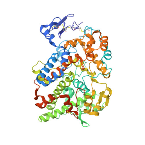
    6V3R

  • PubMed Abstract: 

    We report the design, synthesis, and evaluation of a series of harmaline analogs as selective inhibitors of 2-arachidonylglycerol (2-AG) oxygenation over arachidonic acid (AA) oxygenation by purified cyclooxygenase-2 (COX-2). A fused tricyclic harmaline analog containing a CH 3 O substituent at C-6 and a CH 3 group at the C-1 position of 4,9-dihydro-3 H -pyrido[3,4- b ]indole (compound 3 ) was the best substrate-selective COX-2 inhibitor of those evaluated, exhibiting a 2AG-selective COX-2 inhibitory IC 50 of 0.022 μM as compared to >1 μM for AA. The 2.66 Å resolution crystal complex of COX-2 with compound 3 revealed that this series of tricyclic indoles binds in the cyclooxygenase channel by flipping the side chain of L531 toward the dimer interface. This novel tricyclic indole series provides the foundation for the development of promising substrate-selective molecules capable of increasing endocannabinoid (EC) levels in the brain to offer new treatments for a variety of diseases, from pain and inflammation to stress and anxiety disorders.


  • Organizational Affiliation

    A. B. Hancock, Jr., Memorial Laboratory for Cancer Research, Departments of Biochemistry, Chemistry, and Pharmacology, Vanderbilt Institute of Chemical Biology, and Vanderbilt-Ingram Cancer Center, Vanderbilt University School of Medicine, Nashville, Tennessee 37232, United States.


Macromolecules
Find similar proteins by:  (by identity cutoff)  |  3D Structure
Entity ID: 1
MoleculeChains Sequence LengthOrganismDetailsImage
Prostaglandin G/H synthase 2
A, B, C, D
587Mus musculusMutation(s): 0 
Gene Names: Ptgs2Cox-2Cox2Pghs-bTis10
EC: 1.14.99.1
UniProt
Find proteins for Q05769 (Mus musculus)
Explore Q05769 
Go to UniProtKB:  Q05769
Entity Groups  
Sequence Clusters30% Identity50% Identity70% Identity90% Identity95% Identity100% Identity
UniProt GroupQ05769
Sequence Annotations
Expand
  • Reference Sequence
Oligosaccharides

Help

Entity ID: 2
MoleculeChains Length2D Diagram Glycosylation3D Interactions
2-acetamido-2-deoxy-beta-D-glucopyranose-(1-4)-2-acetamido-2-deoxy-beta-D-glucopyranose
E, F, G, H
2N-Glycosylation
Glycosylation Resources
GlyTouCan:  G42666HT
GlyCosmos:  G42666HT
GlyGen:  G42666HT
Small Molecules
Ligands 4 Unique
IDChains Name / Formula / InChI Key2D Diagram3D Interactions
HEM
Query on HEM

Download Ideal Coordinates CCD File 
I [auth A],
O [auth B],
S [auth C],
W [auth D]
PROTOPORPHYRIN IX CONTAINING FE
C34 H32 Fe N4 O4
KABFMIBPWCXCRK-RGGAHWMASA-L
Y8A (Subject of Investigation/LOI)
Query on Y8A

Download Ideal Coordinates CCD File 
M [auth A],
N [auth B],
R [auth C],
V [auth D]
9-[(4-chlorophenyl)methyl]-6-methoxy-1-methyl-4,9-dihydro-3H-beta-carboline
C20 H19 Cl N2 O
SRMUEFRMPPZYOH-UHFFFAOYSA-N
BOG
Query on BOG

Download Ideal Coordinates CCD File 
L [auth A],
Z [auth D]
octyl beta-D-glucopyranoside
C14 H28 O6
HEGSGKPQLMEBJL-RKQHYHRCSA-N
NAG
Query on NAG

Download Ideal Coordinates CCD File 
J [auth A]
K [auth A]
P [auth B]
Q [auth B]
T [auth C]
J [auth A],
K [auth A],
P [auth B],
Q [auth B],
T [auth C],
U [auth C],
X [auth D],
Y [auth D]
2-acetamido-2-deoxy-beta-D-glucopyranose
C8 H15 N O6
OVRNDRQMDRJTHS-FMDGEEDCSA-N
Experimental Data & Validation

Experimental Data

  • Method: X-RAY DIFFRACTION
  • Resolution: 2.66 Å
  • R-Value Free: 0.264 
  • R-Value Work: 0.219 
  • R-Value Observed: 0.221 
  • Space Group: C 1 2 1
Unit Cell:
Length ( Å )Angle ( ˚ )
a = 217.154α = 90
b = 124.379β = 123.838
c = 136.539γ = 90
Software Package:
Software NamePurpose
PHENIXrefinement
XDSdata reduction
XDSdata scaling
PHASERphasing

Structure Validation

View Full Validation Report

Currently 6V3R does not have a validation slider image.


Entry History & Funding Information

Deposition Data


Funding OrganizationLocationGrant Number
National Institutes of Health/National Cancer Institute (NIH/NCI)United StatesCA89450

Revision History  (Full details and data files)

  • Version 1.0: 2020-02-26
    Type: Initial release
  • Version 2.0: 2020-07-29
    Type: Remediation
    Reason: Carbohydrate remediation
    Changes: Atomic model, Data collection, Derived calculations, Structure summary
  • Version 2.1: 2020-11-04
    Changes: Database references, Structure summary
  • Version 2.2: 2023-10-11
    Changes: Data collection, Database references, Refinement description