


Crystal structure of dihydrofolate reductase from Mycobacterium ulcerans with SDDC-0001565 inhibitor
Mayclin, S.J., Abendroth, J.A., Lorimer, D.D., Horanyi, P.S., Edwards, T.E.To be published.
Experimental Data Snapshot
Starting Model: experimental
View more details
wwPDB Validation 3D Report Full Report
Currently 6UWQ does not have a validation slider image.
Entity ID: 1 | |||||
|---|---|---|---|---|---|
| Molecule | Chains | Sequence Length | Organism | Details | Image |
| Dihydrofolate reductase | 173 | Mycobacterium ulcerans Agy99 | Mutation(s): 0 Gene Names: dfrA, MUL_2179 EC: 1.5.1.3 | 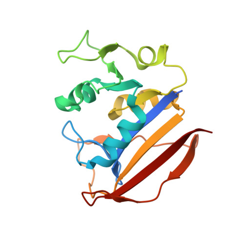 | |
UniProt | |||||
Find proteins for A0PQG8 (Mycobacterium ulcerans (strain Agy99)) Explore A0PQG8 Go to UniProtKB: A0PQG8 | |||||
Entity Groups | |||||
| Sequence Clusters | 30% Identity50% Identity70% Identity90% Identity95% Identity100% Identity | ||||
| UniProt Group | A0PQG8 | ||||
Sequence AnnotationsExpand | |||||
| |||||
| Ligands 3 Unique | |||||
|---|---|---|---|---|---|
| ID | Chains | Name / Formula / InChI Key | 2D Diagram | 3D Interactions | |
| NAP Query on NAP | B [auth A] | NADP NICOTINAMIDE-ADENINE-DINUCLEOTIDE PHOSPHATE C21 H28 N7 O17 P3 XJLXINKUBYWONI-NNYOXOHSSA-N |  | ||
| QKJ (Subject of Investigation/LOI) Query on QKJ | C [auth A] | 3-(3-{3-[(2,4-diamino-6-ethylpyrimidin-5-yl)oxy]propoxy}phenyl)propanoic acid C18 H24 N4 O4 HMQJRDLOETYJSB-UHFFFAOYSA-N |  | ||
| EDO Query on EDO | D [auth A], E [auth A] | 1,2-ETHANEDIOL C2 H6 O2 LYCAIKOWRPUZTN-UHFFFAOYSA-N |  | ||
| Length ( Å ) | Angle ( ˚ ) |
|---|---|
| a = 29.29 | α = 90 |
| b = 70.75 | β = 110.991 |
| c = 35.99 | γ = 90 |
| Software Name | Purpose |
|---|---|
| XDS | data reduction |
| XSCALE | data scaling |
| PHASER | phasing |
| PHENIX | refinement |
| PDB_EXTRACT | data extraction |
Currently 6UWQ does not have a validation slider image.
RCSB PDB (citation) is hosted by
RCSB PDB is a member of the

