6UIU

Artificial Iron Proteins: Modelling the Active Sites in Non-Heme Dioxygenases


Experimental Data Snapshot

  • Method: X-RAY DIFFRACTION
  • Resolution: 1.35 Å
  • R-Value Free: 0.230 
  • R-Value Work: 0.207 
  • R-Value Observed: 0.208 

wwPDB Validation   3D Report Full Report


Ligand Structure Quality Assessment 


This is version 1.2 of the entry. See complete history


Literature

Artificial Iron Proteins: Modeling the Active Sites in Non-Heme Dioxygenases.

Miller, K.R.Paretsky, J.D.Follmer, A.H.Heinisch, T.Mittra, K.Gul, S.Kim, I.S.Fuller, F.D.Batyuk, A.Sutherlin, K.D.Brewster, A.S.Bhowmick, A.Sauter, N.K.Kern, J.Yano, J.Green, M.T.Ward, T.R.Borovik, A.S.

(2020) Inorg Chem 59: 6000-6009

  • DOI: https://doi.org/10.1021/acs.inorgchem.9b03791
  • Primary Citation of Related Structures:  
    6UI0, 6UIU, 6UIY, 6UIZ, 6US6

  • PubMed Abstract: 

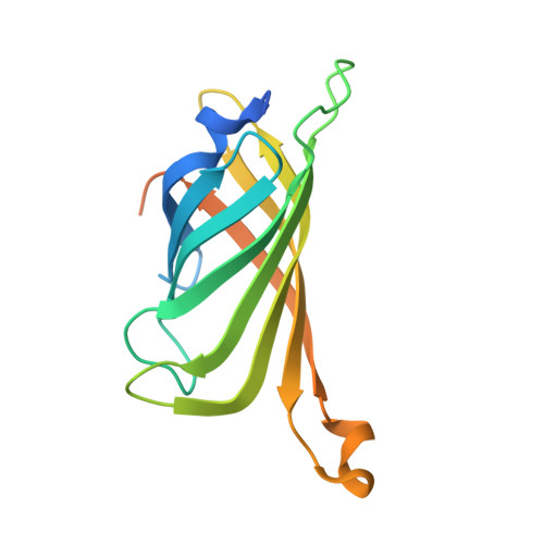
    An important class of non-heme dioxygenases contains a conserved Fe binding site that consists of a 2-His-1-carboxylate facial triad. Results from structural biology show that, in the resting state, these proteins are six-coordinate with aqua ligands occupying the remaining three coordination sites. We have utilized biotin-streptavidin (Sav) technology to design new artificial Fe proteins (ArMs) that have many of the same structural features found within active sites of these non-heme dioxygenases. An Sav variant was isolated that contains the S 112 E mutation, which installed a carboxylate side chain in the appropriate position to bind to a synthetic Fe II complex confined within Sav. Structural studies using X-ray diffraction (XRD) methods revealed a facial triad binding site that is composed of two N donors from the biotinylated ligand and the monodentate coordination of the carboxylate from S 112 E. Two aqua ligands complete the primary coordination sphere of the Fe II center with both involved in hydrogen bond networks within Sav. The corresponding Fe III protein was also prepared and structurally characterized to show a six-coordinate complex with two exogenous acetato ligands. The Fe III protein was further shown to bind an exogenous azido ligand through replacement of one acetato ligand. Spectroscopic studies of the ArMs in solution support the results found by XRD.


  • Organizational Affiliation

    Department of Chemistry, 1102 Natural Science II, University of California, Irvine, California 92697, United States.


Macromolecules
Find similar proteins by:  (by identity cutoff)  |  3D Structure
Entity ID: 1
MoleculeChains Sequence LengthOrganismDetailsImage
Streptavidin159Streptomyces avidiniiMutation(s): 3 
UniProt
Find proteins for P22629 (Streptomyces avidinii)
Explore P22629 
Go to UniProtKB:  P22629
Entity Groups  
Sequence Clusters30% Identity50% Identity70% Identity90% Identity95% Identity100% Identity
UniProt GroupP22629
Sequence Annotations
Expand
  • Reference Sequence
Small Molecules
Ligands 1 Unique
IDChains Name / Formula / InChI Key2D Diagram3D Interactions
QG7 (Subject of Investigation/LOI)
Query on QG7

Download Ideal Coordinates CCD File 
B [auth A]N-(2-{bis[(pyridin-2-yl)methyl]amino}ethyl)-5-[(3aS,4S,6aR)-2-oxohexahydro-1H-thieno[3,4-d]imidazol-4-yl]pentanamide
C24 H32 N6 O2 S
GXLJCVSDSFWSLD-FUDKSRODSA-N
Experimental Data & Validation

Experimental Data

  • Method: X-RAY DIFFRACTION
  • Resolution: 1.35 Å
  • R-Value Free: 0.230 
  • R-Value Work: 0.207 
  • R-Value Observed: 0.208 
  • Space Group: I 41 2 2
Unit Cell:
Length ( Å )Angle ( ˚ )
a = 57.871α = 90
b = 57.871β = 90
c = 184.305γ = 90
Software Package:
Software NamePurpose
XDSdata reduction
Aimlessdata scaling
PHASERphasing
REFMACrefinement
PDB_EXTRACTdata extraction

Structure Validation

View Full Validation Report


Ligand Structure Quality Assessment 


Entry History & Funding Information

Deposition Data


Funding OrganizationLocationGrant Number
National Institutes of Health/National Institute of General Medical Sciences (NIH/NIGMS)United StatesRO1GM120349

Revision History  (Full details and data files)

  • Version 1.0: 2020-05-06
    Type: Initial release
  • Version 1.1: 2020-05-13
    Changes: Database references
  • Version 1.2: 2023-10-11
    Changes: Data collection, Database references, Refinement description