6UIO

Crystal structure of mouse CRES (Cystatin-Related Epididymal Spermatogenic)


Experimental Data Snapshot

  • Method: X-RAY DIFFRACTION
  • Resolution: 1.83 Å
  • R-Value Free: 0.262 
  • R-Value Work: 0.224 
  • R-Value Observed: 0.228 

wwPDB Validation   3D Report Full Report


This is version 1.3 of the entry. See complete history


Literature

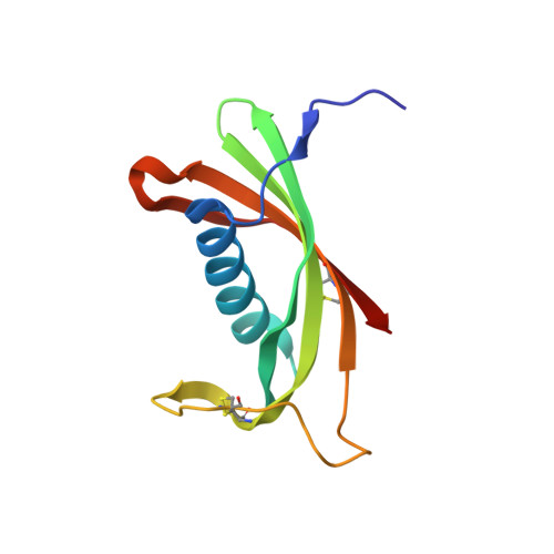
Maturation of the functional mouse CRES amyloid from globular form.

Hewetson, A.Khan, N.H.Dominguez, M.J.Do, H.Q.Kusko, R.E.Borcik, C.G.Rigden, D.J.Keegan, R.M.Sutton, R.B.Latham, M.P.Wylie, B.J.Cornwall, G.A.

(2020) Proc Natl Acad Sci U S A 117: 16363-16372

  • DOI: https://doi.org/10.1073/pnas.2006887117
  • Primary Citation of Related Structures:  
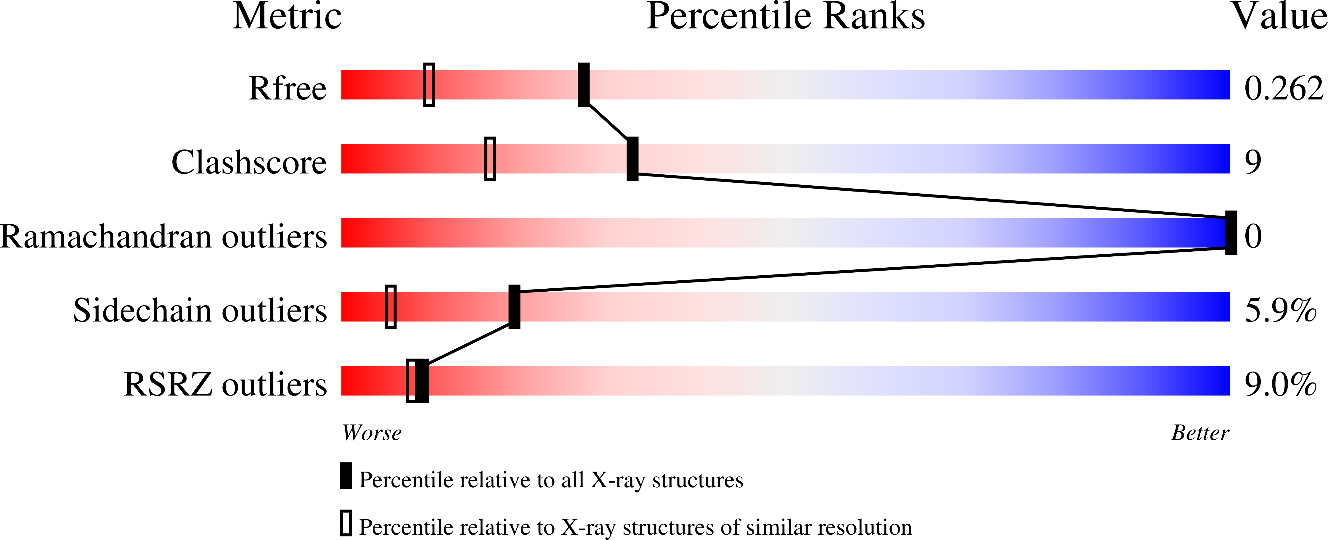
    6UIO

  • PubMed Abstract: 

    The epididymal lumen contains a complex cystatin-rich nonpathological amyloid matrix with putative roles in sperm maturation and sperm protection. Given our growing understanding for the biological function of this and other functional amyloids, the problem still remains: how functional amyloids assemble including their initial transition to early oligomeric forms. To examine this, we developed a protocol for the purification of nondenatured mouse CRES, a component of the epididymal amyloid matrix, allowing us to examine its assembly to amyloid under conditions that may mimic those in vivo. Herein we use X-ray crystallography, solution-state NMR, and solid-state NMR to follow at the atomic level the assembly of the CRES amyloidogenic precursor as it progressed from monomeric folded protein to an advanced amyloid. We show the CRES monomer has a typical cystatin fold that assembles into highly branched amyloid matrices, comparable to those in vivo, by forming β-sheet assemblies that our data suggest occur via two distinct mechanisms: a unique conformational switch of a highly flexible disulfide-anchored loop to a rigid β-strand and by traditional cystatin domain swapping. Our results provide key insight into our understanding of functional amyloid assembly by revealing the earliest structural transitions from monomer to oligomer and by showing that some functional amyloid structures may be built by multiple and distinctive assembly mechanisms.


  • Organizational Affiliation

    Department of Cell Biology and Biochemistry, Texas Tech University Health Sciences Center, Lubbock, TX 79430.


Macromolecules
Find similar proteins by:  (by identity cutoff)  |  3D Structure
Entity ID: 1
MoleculeChains Sequence LengthOrganismDetailsImage
Cystatin-8
A, B, C, D
112Mus musculusMutation(s): 1 
Gene Names: Cst8Cres
UniProt
Find proteins for P32766 (Mus musculus)
Explore P32766 
Go to UniProtKB:  P32766
Entity Groups  
Sequence Clusters30% Identity50% Identity70% Identity90% Identity95% Identity100% Identity
UniProt GroupP32766
Sequence Annotations
Expand
  • Reference Sequence
Small Molecules
Ligands 6 Unique
IDChains Name / Formula / InChI Key2D Diagram3D Interactions
I2I
Query on I2I

Download Ideal Coordinates CCD File 
R [auth B],
ZA [auth D]
molecular iodine
I2
PNDPGZBMCMUPRI-UHFFFAOYSA-N
MES
Query on MES

Download Ideal Coordinates CCD File 
FB [auth D],
OA [auth C]
2-(N-MORPHOLINO)-ETHANESULFONIC ACID
C6 H13 N O4 S
SXGZJKUKBWWHRA-UHFFFAOYSA-N
IOD
Query on IOD

Download Ideal Coordinates CCD File 
BA [auth C]
CA [auth C]
DA [auth C]
E [auth A]
EA [auth C]
BA [auth C],
CA [auth C],
DA [auth C],
E [auth A],
EA [auth C],
F [auth A],
FA [auth C],
G [auth A],
GA [auth C],
H [auth A],
HA [auth C],
I [auth A],
P [auth B],
Q [auth B],
RA [auth D],
SA [auth D],
TA [auth D],
UA [auth D],
VA [auth D],
WA [auth D],
XA [auth D],
YA [auth D]
IODIDE ION
I
XMBWDFGMSWQBCA-UHFFFAOYSA-M
SO4
Query on SO4

Download Ideal Coordinates CCD File 
AB [auth D]
BB [auth D]
CB [auth D]
IA [auth C]
J [auth A]
AB [auth D],
BB [auth D],
CB [auth D],
IA [auth C],
J [auth A],
JA [auth C],
K [auth A],
KA [auth C],
L [auth A],
M [auth A],
N [auth A],
O [auth A],
S [auth B],
T [auth B],
U [auth B],
V [auth B],
W [auth B],
X [auth B]
SULFATE ION
O4 S
QAOWNCQODCNURD-UHFFFAOYSA-L
CL
Query on CL

Download Ideal Coordinates CCD File 
AA [auth B]
DB [auth D]
EB [auth D]
LA [auth C]
MA [auth C]
AA [auth B],
DB [auth D],
EB [auth D],
LA [auth C],
MA [auth C],
NA [auth C],
Y [auth B],
Z [auth B]
CHLORIDE ION
Cl
VEXZGXHMUGYJMC-UHFFFAOYSA-M
NA
Query on NA

Download Ideal Coordinates CCD File 
GB [auth D]
HB [auth D]
IB [auth D]
JB [auth D]
PA [auth C]
GB [auth D],
HB [auth D],
IB [auth D],
JB [auth D],
PA [auth C],
QA [auth C]
SODIUM ION
Na
FKNQFGJONOIPTF-UHFFFAOYSA-N
Experimental Data & Validation

Experimental Data

  • Method: X-RAY DIFFRACTION
  • Resolution: 1.83 Å
  • R-Value Free: 0.262 
  • R-Value Work: 0.224 
  • R-Value Observed: 0.228 
  • Space Group: P 21 21 21
Unit Cell:
Length ( Å )Angle ( ˚ )
a = 51.048α = 90
b = 85.924β = 90
c = 111.677γ = 90
Software Package:
Software NamePurpose
PHENIXrefinement
HKL-3000data reduction
SCALEPACKdata scaling
PHASERphasing

Structure Validation

View Full Validation Report


Entry History 

Deposition Data

Revision History  (Full details and data files)

  • Version 1.0: 2020-07-01
    Type: Initial release
  • Version 1.1: 2020-07-15
    Changes: Database references
  • Version 1.2: 2020-07-29
    Changes: Database references, Derived calculations
  • Version 1.3: 2023-10-11
    Changes: Data collection, Database references, Refinement description