6U9V

Cryo electron microscopy structure of the ATP-gated rat P2X7 ion channel in the apo, closed state


Experimental Data Snapshot

  • Method: ELECTRON MICROSCOPY
  • Resolution: 2.90 Å
  • Aggregation State: PARTICLE 
  • Reconstruction Method: SINGLE PARTICLE 

wwPDB Validation   3D Report Full Report


This is version 2.0 of the entry. See complete history


Literature

Full-Length P2X7Structures Reveal How Palmitoylation Prevents Channel Desensitization.

McCarthy, A.E.Yoshioka, C.Mansoor, S.E.

(2019) Cell 179: 659-670.e13

  • DOI: https://doi.org/10.1016/j.cell.2019.09.017
  • Primary Citation of Related Structures:  
    6U9V, 6U9W

  • PubMed Abstract: 

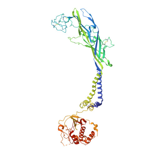
    P2X receptors are trimeric, non-selective cation channels activated by extracellular ATP. The P2X 7 receptor subtype is a pharmacological target because of involvement in apoptotic, inflammatory, and tumor progression pathways. It is the most structurally and functionally distinct P2X subtype, containing a unique cytoplasmic domain critical for the receptor to initiate apoptosis and not undergo desensitization. However, lack of structural information about the cytoplasmic domain has hindered understanding of the molecular mechanisms underlying these processes. We report cryoelectron microscopy structures of full-length rat P2X 7 receptor in apo and ATP-bound states. These structures reveal how one cytoplasmic element, the C-cys anchor, prevents desensitization by anchoring the pore-lining helix to the membrane with palmitoyl groups. They show a second cytoplasmic element with a unique fold, the cytoplasmic ballast, which unexpectedly contains a zinc ion complex and a guanosine nucleotide binding site. Our structures provide first insights into the architecture and function of a P2X receptor cytoplasmic domain.


  • Organizational Affiliation

    Vollum Institute, Oregon Health and Science University, Portland, OR 97239, USA; Knight Cardiovascular Institute, Oregon Health and Science University, Portland, OR 97239, USA.


Macromolecules
Find similar proteins by:  (by identity cutoff)  |  3D Structure
Entity ID: 1
MoleculeChains Sequence LengthOrganismDetailsImage
P2X purinoceptor 7
A, B, C
609Rattus norvegicusMutation(s): 0 
Gene Names: P2rx7
Membrane Entity: Yes 
UniProt
Find proteins for Q64663 (Rattus norvegicus)
Explore Q64663 
Go to UniProtKB:  Q64663
Entity Groups  
Sequence Clusters30% Identity50% Identity70% Identity90% Identity95% Identity100% Identity
UniProt GroupQ64663
Sequence Annotations
Expand
  • Reference Sequence
Oligosaccharides

Help

Entity ID: 2
MoleculeChains Length2D Diagram Glycosylation3D Interactions
2-acetamido-2-deoxy-beta-D-glucopyranose-(1-4)-2-acetamido-2-deoxy-beta-D-glucopyranose
D, E, F, G, H
D, E, F, G, H, I
2N-Glycosylation
Glycosylation Resources
GlyTouCan:  G42666HT
GlyCosmos:  G42666HT
GlyGen:  G42666HT
Entity ID: 3
MoleculeChains Length2D Diagram Glycosylation3D Interactions
alpha-D-glucopyranose-(1-4)-alpha-D-glucopyranose
J, K, L
2N/A
Glycosylation Resources
GlyTouCan:  G07411ON
GlyCosmos:  G07411ON
Small Molecules
Ligands 5 Unique
IDChains Name / Formula / InChI Key2D Diagram3D Interactions
Q3G
Query on Q3G

Download Ideal Coordinates CCD File 
FA [auth B],
S [auth A],
SA [auth C]
O-[(R)-[(2S)-2-(hexadecanoyloxy)-3-(octadecanoyloxy)propoxy](hydroxy)phosphoryl]-D-serine
C40 H78 N O10 P
WZFUPCSEUKNOBF-PQQNNWGCSA-N
GDP (Subject of Investigation/LOI)
Query on GDP

Download Ideal Coordinates CCD File 
M [auth A],
MA [auth C],
Z [auth B]
GUANOSINE-5'-DIPHOSPHATE
C10 H15 N5 O11 P2
QGWNDRXFNXRZMB-UUOKFMHZSA-N
PLM
Query on PLM

Download Ideal Coordinates CCD File 
GA [auth B]
HA [auth B]
IA [auth B]
JA [auth B]
KA [auth B]
GA [auth B],
HA [auth B],
IA [auth B],
JA [auth B],
KA [auth B],
LA [auth B],
T [auth A],
TA [auth C],
U [auth A],
UA [auth C],
V [auth A],
VA [auth C],
W [auth A],
WA [auth C],
X [auth A],
XA [auth C],
Y [auth A],
YA [auth C]
PALMITIC ACID
C16 H32 O2
IPCSVZSSVZVIGE-UHFFFAOYSA-N
NAG
Query on NAG

Download Ideal Coordinates CCD File 
CA [auth B]
DA [auth B]
EA [auth B]
P [auth A]
PA [auth C]
CA [auth B],
DA [auth B],
EA [auth B],
P [auth A],
PA [auth C],
Q [auth A],
QA [auth C],
R [auth A],
RA [auth C]
2-acetamido-2-deoxy-beta-D-glucopyranose
C8 H15 N O6
OVRNDRQMDRJTHS-FMDGEEDCSA-N
ZN (Subject of Investigation/LOI)
Query on ZN

Download Ideal Coordinates CCD File 
AA [auth B]
BA [auth B]
N [auth A]
NA [auth C]
O [auth A]
AA [auth B],
BA [auth B],
N [auth A],
NA [auth C],
O [auth A],
OA [auth C]
ZINC ION
Zn
PTFCDOFLOPIGGS-UHFFFAOYSA-N
Biologically Interesting Molecules (External Reference) 1 Unique
Experimental Data & Validation

Experimental Data

  • Method: ELECTRON MICROSCOPY
  • Resolution: 2.90 Å
  • Aggregation State: PARTICLE 
  • Reconstruction Method: SINGLE PARTICLE 

Structure Validation

View Full Validation Report


Entry History & Funding Information

Deposition Data


Funding OrganizationLocationGrant Number
National Institutes of Health/National Heart, Lung, and Blood Institute (NIH/NHLBI)United StatesK99HL138129

Revision History  (Full details and data files)

  • Version 1.0: 2019-10-23
    Type: Initial release
  • Version 1.1: 2019-10-30
    Changes: Data collection, Database references
  • Version 1.2: 2019-12-04
    Changes: Author supporting evidence
  • Version 2.0: 2020-07-29
    Type: Remediation
    Reason: Carbohydrate remediation
    Changes: Advisory, Atomic model, Data collection, Derived calculations, Non-polymer description, Structure summary