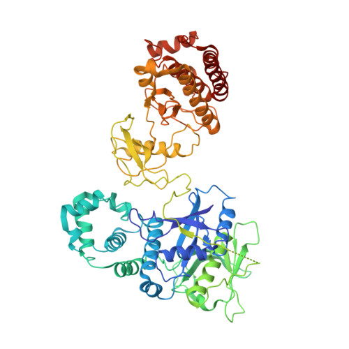
Structural basis of Focal Adhesion Kinase activation on lipid membranes.
Acebron, I., Righetto, R.D., Schoenherr, C., de Buhr, S., Redondo, P., Culley, J., Rodriguez, C.F., Daday, C., Biyani, N., Llorca, O., Byron, A., Chami, M., Grater, F., Boskovic, J., Frame, M.C., Stahlberg, H., Lietha, D.(2020) EMBO J 39: e104743-e104743
- PubMed: 32779739
- DOI: https://doi.org/10.15252/embj.2020104743
- Primary Citation of Related Structures:
6TY3, 6TY4 - PubMed Abstract:
Focal adhesion kinase (FAK) is a key component of the membrane proximal signaling layer in focal adhesion complexes, regulating important cellular processes, including cell migration, proliferation, and survival. In the cytosol, FAK adopts an autoinhibited state but is activated upon recruitment into focal adhesions, yet how this occurs or what induces structural changes is unknown. Here, we employ cryo-electron microscopy to reveal how FAK associates with lipid membranes and how membrane interactions unlock FAK autoinhibition to promote activation. Intriguingly, initial binding of FAK to the membrane causes steric clashes that release the kinase domain from autoinhibition, allowing it to undergo a large conformational change and interact itself with the membrane in an orientation that places the active site toward the membrane. In this conformation, the autophosphorylation site is exposed and multiple interfaces align to promote FAK oligomerization on the membrane. We show that interfaces responsible for initial dimerization and membrane attachment are essential for FAK autophosphorylation and resulting cellular activity including cancer cell invasion, while stable FAK oligomerization appears to be needed for optimal cancer cell proliferation in an anchorage-independent manner. Together, our data provide structural details of a key membrane bound state of FAK that is primed for efficient autophosphorylation and activation, hence revealing the critical event in integrin mediated FAK activation and signaling at focal adhesions.
Organizational Affiliation:
Structural Biology Programme, Spanish National Cancer Research Centre, Madrid, Spain.