6TTG

Crystal structure of the ATP binding domain of S. aureus GyrB complexed with LMD62


Experimental Data Snapshot

  • Method: X-RAY DIFFRACTION
  • Resolution: 1.70 Å
  • R-Value Free: 0.211 
  • R-Value Work: 0.186 
  • R-Value Observed: 0.187 

wwPDB Validation   3D Report Full Report


Ligand Structure Quality Assessment 


This is version 1.2 of the entry. See complete history


Literature

New dual ATP-competitive inhibitors of bacterial DNA gyrase and topoisomerase IV active against ESKAPE pathogens.

Durcik, M.Nyerges, A.Skok, Z.Skledar, D.G.Trontelj, J.Zidar, N.Ilas, J.Zega, A.Cruz, C.D.Tammela, P.Welin, M.Kimbung, Y.R.Focht, D.Benek, O.Revesz, T.Draskovits, G.Szili, P.E.Daruka, L.Pal, C.Kikelj, D.Masic, L.P.Tomasic, T.

(2021) Eur J Med Chem 213: 113200-113200

  • DOI: https://doi.org/10.1016/j.ejmech.2021.113200
  • Primary Citation of Related Structures:  
    6TTG

  • PubMed Abstract: 

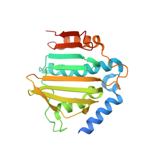
    The rise in multidrug-resistant bacteria defines the need for identification of new antibacterial agents that are less prone to resistance acquisition. Compounds that simultaneously inhibit multiple bacterial targets are more likely to suppress the evolution of target-based resistance than monotargeting compounds. The structurally similar ATP binding sites of DNA gyrase and topoisomerase Ⅳ offer an opportunity to accomplish this goal. Here we present the design and structure-activity relationship analysis of balanced, low nanomolar inhibitors of bacterial DNA gyrase and topoisomerase IV that show potent antibacterial activities against the ESKAPE pathogens. For inhibitor 31c, a crystal structure in complex with Staphylococcus aureus DNA gyrase B was obtained that confirms the mode of action of these compounds. The best inhibitor, 31h, does not show any in vitro cytotoxicity and has excellent potency against Gram-positive (MICs: range, 0.0078-0.0625 μg/mL) and Gram-negative pathogens (MICs: range, 1-2 μg/mL). Furthermore, 31h inhibits GyrB mutants that can develop resistance to other drugs. Based on these data, we expect that structural derivatives of 31h will represent a step toward clinically efficacious multitargeting antimicrobials that are not impacted by existing antimicrobial resistance.


  • Organizational Affiliation

    University of Ljubljana, Faculty of Pharmacy, Aškerčeva cesta 7, 1000, Ljubljana, Slovenia.


Macromolecules
Find similar proteins by:  (by identity cutoff)  |  3D Structure
Entity ID: 1
MoleculeChains Sequence LengthOrganismDetailsImage
DNA gyrase subunit B
A, B
212Staphylococcus aureusMutation(s): 0 
Gene Names: gyrB
EC: 5.6.2.2
UniProt
Find proteins for P0A0K8 (Staphylococcus aureus)
Explore P0A0K8 
Go to UniProtKB:  P0A0K8
Entity Groups  
Sequence Clusters30% Identity50% Identity70% Identity90% Identity95% Identity100% Identity
UniProt GroupP0A0K8
Sequence Annotations
Expand
  • Reference Sequence
Small Molecules
Ligands 6 Unique
IDChains Name / Formula / InChI Key2D Diagram3D Interactions
NWK (Subject of Investigation/LOI)
Query on NWK

Download Ideal Coordinates CCD File 
C [auth A],
J [auth B]
2-[[3,4-bis(chloranyl)-5-methyl-1~{H}-pyrrol-2-yl]carbonylamino]-4-(2-morpholin-4-ylethoxy)-1,3-benzothiazole-6-carboxylic acid
C20 H20 Cl2 N4 O5 S
IALGYKMHQTUZKU-UHFFFAOYSA-N
MPD
Query on MPD

Download Ideal Coordinates CCD File 
E [auth A],
F [auth A],
G [auth A],
K [auth B],
L [auth B]
(4S)-2-METHYL-2,4-PENTANEDIOL
C6 H14 O2
SVTBMSDMJJWYQN-YFKPBYRVSA-N
GOL
Query on GOL

Download Ideal Coordinates CCD File 
D [auth A]GLYCEROL
C3 H8 O3
PEDCQBHIVMGVHV-UHFFFAOYSA-N
IMD
Query on IMD

Download Ideal Coordinates CCD File 
M [auth B]IMIDAZOLE
C3 H5 N2
RAXXELZNTBOGNW-UHFFFAOYSA-O
CA
Query on CA

Download Ideal Coordinates CCD File 
H [auth A]CALCIUM ION
Ca
BHPQYMZQTOCNFJ-UHFFFAOYSA-N
CL
Query on CL

Download Ideal Coordinates CCD File 
I [auth A],
N [auth B]
CHLORIDE ION
Cl
VEXZGXHMUGYJMC-UHFFFAOYSA-M
Experimental Data & Validation

Experimental Data

  • Method: X-RAY DIFFRACTION
  • Resolution: 1.70 Å
  • R-Value Free: 0.211 
  • R-Value Work: 0.186 
  • R-Value Observed: 0.187 
  • Space Group: C 1 2 1
Unit Cell:
Length ( Å )Angle ( ˚ )
a = 142.7α = 90
b = 55.36β = 100.42
c = 50.81γ = 90
Software Package:
Software NamePurpose
REFMACrefinement
PDB_EXTRACTdata extraction
xia2data reduction
xia2data scaling
REFMACphasing

Structure Validation

View Full Validation Report


Ligand Structure Quality Assessment 


Entry History & Funding Information

Deposition Data


Funding OrganizationLocationGrant Number
Slovenian Research AgencySloveniaP1-0208

Revision History  (Full details and data files)

  • Version 1.0: 2020-12-30
    Type: Initial release
  • Version 1.1: 2022-01-26
    Changes: Database references
  • Version 1.2: 2024-01-24
    Changes: Data collection, Refinement description