6TT9

rTBL Recombinant Lectin From Tepary Bean


Experimental Data Snapshot

  • Method: X-RAY DIFFRACTION
  • Resolution: 1.90 Å
  • R-Value Free: 0.213 
  • R-Value Work: 0.169 

wwPDB Validation   3D Report Full Report


This is version 2.1 of the entry. See complete history


Literature

Recombinant Lectin from Tepary Bean (Phaseolus acutifolius) with Specific Recognition for Cancer-Associated Glycans: Production, Structural Characterization, and Target Identification.

Martinez-Alarcon, D.Varrot, A.Fitches, E.Gatehouse, J.A.Cao, M.Pyati, P.Blanco-Labra, A.Garcia-Gasca, T.

(2020) Biomolecules 10

  • DOI: https://doi.org/10.3390/biom10040654
  • Primary Citation of Related Structures:  
    6TT9

  • PubMed Abstract: 

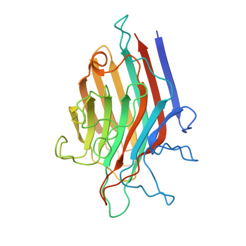
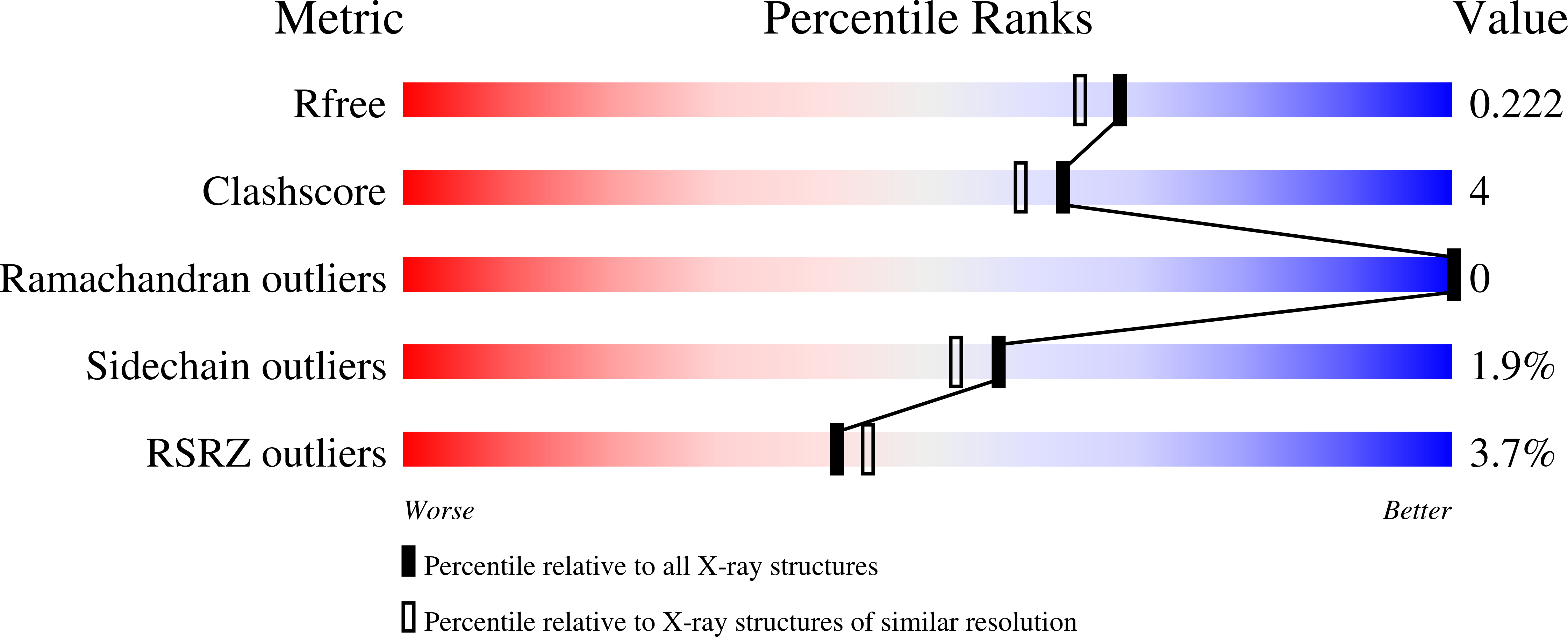
    Herein, we report the production of a recombinant Tepary bean lectin ( r TBL-1), its three-dimensional (3D) structure, and its differential recognition for cancer-type glycoconjugates. r TBL-1 was expressed in Pichia pastoris, yielding 316 mg per liter of culture, and was purified by nickel affinity chromatography. Characterization of the protein showed that r TBL-1 is a stable 120 kDa homo-tetramer folded as a canonical leguminous lectin with two divalent cations (Ca 2+ and Mn 2+ ) attached to each subunit, confirmed in its 3D structure solved by X-ray diffraction at 1.9 Å resolution. Monomers also presented a ~2.5 kDa N -linked glycan located on the opposite face of the binding pocket. It does not participate in carbohydrate recognition but contributes to the stabilization of the interfaces between protomers. Screening for potential r TBL-1 targets by glycan array identified 14 positive binders, all of which correspond to β1-6 branched N -glycans' characteristics of cancer cells. The presence of α1-6 core fucose, also tumor-associated, improved carbohydrate recognition. r TBL-1 affinity for a broad spectrum of mono- and disaccharides was evaluated by isothermal titration calorimetry (ITC); however, no interaction was detected, corroborating that carbohydrate recognition is highly specific and requires larger ligands for binding. This would explain the differential recognition between healthy and cancer cells by Tepary bean lectins.


  • Organizational Affiliation

    Centro de Investigación y de Estudios Avanzados Unidad Irapuato, Departamento de Biotecnología y Bioquímica, Irapuato 36821, Guanaj uato, Mexico.


Macromolecules
Find similar proteins by:  (by identity cutoff)  |  3D Structure
Entity ID: 1
MoleculeChains Sequence LengthOrganismDetailsImage
PhytohemagglutininA [auth AAA],
B [auth BBB],
C [auth CCC],
D [auth DDD]
255Phaseolus acutifoliusMutation(s): 0 
UniProt
Find proteins for Q40750 (Phaseolus acutifolius)
Explore Q40750 
Go to UniProtKB:  Q40750
Entity Groups  
Sequence Clusters30% Identity50% Identity70% Identity90% Identity95% Identity100% Identity
UniProt GroupQ40750
Sequence Annotations
Expand
  • Reference Sequence
Oligosaccharides

Help

Entity ID: 2
MoleculeChains Length2D Diagram Glycosylation3D Interactions
alpha-D-mannopyranose-(1-2)-alpha-D-mannopyranose-(1-6)-[alpha-D-mannopyranose-(1-3)]alpha-D-mannopyranose-(1-6)-[alpha-D-mannopyranose-(1-3)]beta-D-mannopyranose-(1-4)-2-acetamido-2-deoxy-beta-D-glucopyranose-(1-4)-2-acetamido-2-deoxy-beta-D-glucopyranoseE [auth A]8N-Glycosylation
Glycosylation Resources
GlyTouCan:  G61846BY
GlyCosmos:  G61846BY
GlyGen:  G61846BY
Entity ID: 3
MoleculeChains Length2D Diagram Glycosylation3D Interactions
2-acetamido-2-deoxy-beta-D-glucopyranose-(1-4)-2-acetamido-2-deoxy-beta-D-glucopyranoseF [auth B],
G [auth C]
2N-Glycosylation
Glycosylation Resources
GlyTouCan:  G42666HT
GlyCosmos:  G42666HT
GlyGen:  G42666HT
Entity ID: 4
MoleculeChains Length2D Diagram Glycosylation3D Interactions
alpha-D-mannopyranose-(1-2)-alpha-D-mannopyranose-(1-6)-alpha-D-mannopyranose-(1-6)-beta-D-mannopyranose-(1-4)-2-acetamido-2-deoxy-beta-D-glucopyranose-(1-4)-2-acetamido-2-deoxy-beta-D-glucopyranoseH [auth D]6N-Glycosylation
Glycosylation Resources
GlyTouCan:  G02763QD
GlyCosmos:  G02763QD
GlyGen:  G02763QD
Small Molecules
Ligands 3 Unique
IDChains Name / Formula / InChI Key2D Diagram3D Interactions
HEX
Query on HEX

Download Ideal Coordinates CCD File 
K [auth AAA],
N [auth BBB],
Q [auth CCC],
R [auth CCC],
U [auth DDD]
HEXANE
C6 H14
VLKZOEOYAKHREP-UHFFFAOYSA-N
MN
Query on MN

Download Ideal Coordinates CCD File 
J [auth AAA],
M [auth BBB],
P [auth CCC],
T [auth DDD]
MANGANESE (II) ION
Mn
WAEMQWOKJMHJLA-UHFFFAOYSA-N
CA
Query on CA

Download Ideal Coordinates CCD File 
I [auth AAA],
L [auth BBB],
O [auth CCC],
S [auth DDD]
CALCIUM ION
Ca
BHPQYMZQTOCNFJ-UHFFFAOYSA-N
Experimental Data & Validation

Experimental Data

  • Method: X-RAY DIFFRACTION
  • Resolution: 1.90 Å
  • R-Value Free: 0.213 
  • R-Value Work: 0.169 
  • Space Group: P 1
Unit Cell:
Length ( Å )Angle ( ˚ )
a = 57.572α = 96.352
b = 64.472β = 101.952
c = 67.59γ = 97.268
Software Package:
Software NamePurpose
REFMACrefinement
XDSdata reduction
Aimlessdata scaling
PHASERphasing

Structure Validation

View Full Validation Report


Entry History & Funding Information

Deposition Data


Funding OrganizationLocationGrant Number
Consejo Nacional de Ciencia y Tecnologia (CONACYT)MexicoCB-2014-01-241181
European CommissionFrance765581

Revision History  (Full details and data files)

  • Version 1.0: 2020-05-13
    Type: Initial release
  • Version 2.0: 2020-07-29
    Type: Remediation
    Reason: Carbohydrate remediation
    Changes: Atomic model, Data collection, Derived calculations, Structure summary
  • Version 2.1: 2024-01-24
    Changes: Data collection, Database references, Derived calculations, Refinement description, Structure summary