6TM3

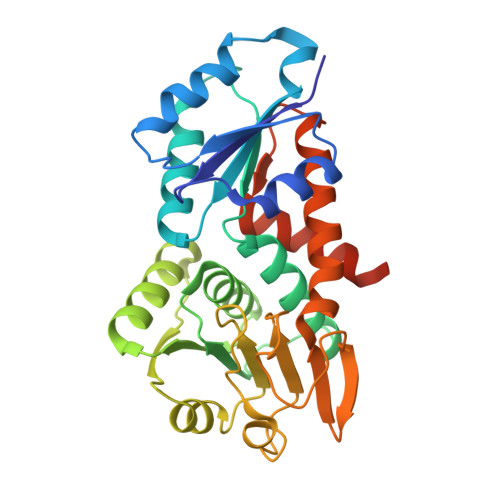
Structure of methylene-tetrahydromethanopterin dehydrogenase from Methylorubrum extorquens AM1 in a close conformation containing NADP+ and methylene-H4MPT


Experimental Data Snapshot

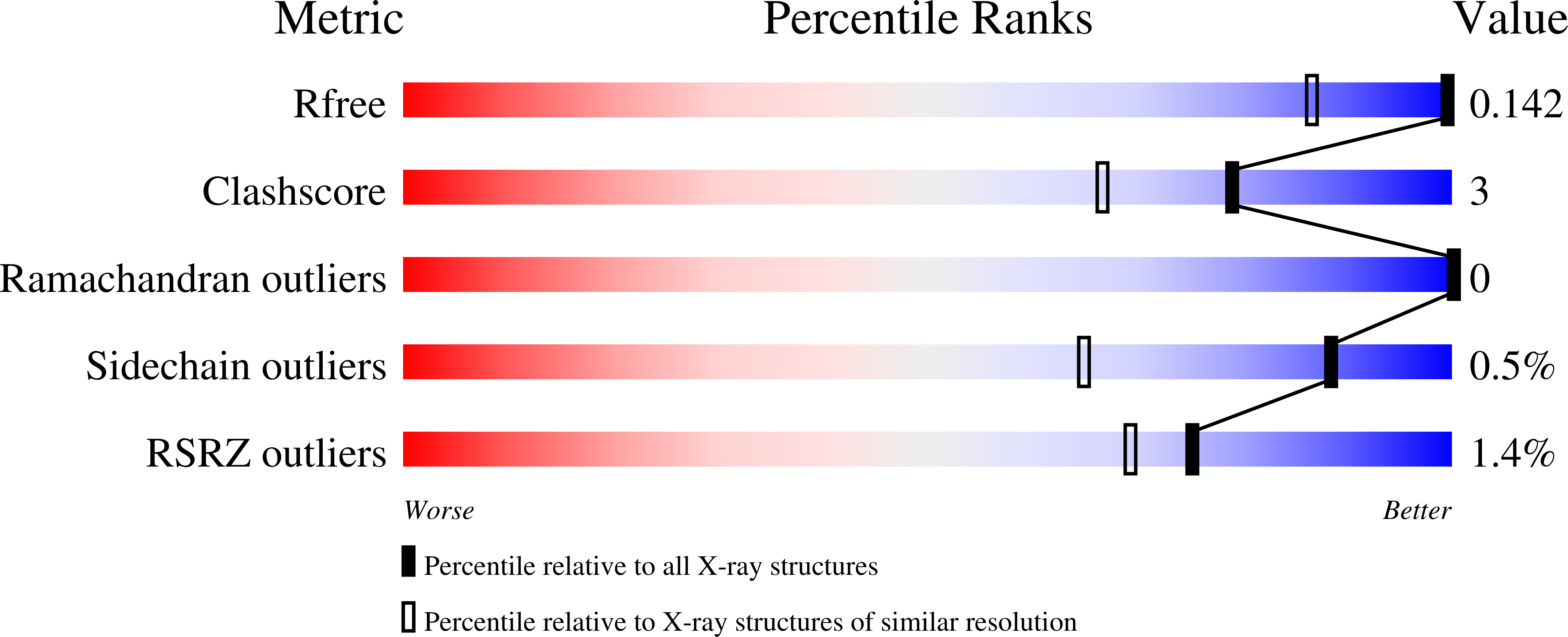
  • Method: X-RAY DIFFRACTION
  • Resolution: 1.08 Å
  • R-Value Free: 0.127 
  • R-Value Work: 0.113 
  • R-Value Observed: 0.114 

wwPDB Validation   3D Report Full Report


Ligand Structure Quality Assessment 


This is version 1.2 of the entry. See complete history


Literature

The Hydride Transfer Process in NADP-dependent Methylene-tetrahydromethanopterin Dehydrogenase.

Huang, G.Wagner, T.Demmer, U.Warkentin, E.Ermler, U.Shima, S.

(2020) J Mol Biol 432: 2042-2054

  • DOI: https://doi.org/10.1016/j.jmb.2020.01.042
  • Primary Citation of Related Structures:  
    6TGE, 6TLK, 6TM3, 6YK9, 6YKA, 6YKB

  • PubMed Abstract: 

    NADP-dependent methylene-tetrahydromethanopterin (methylene-H 4 MPT) dehydrogenase (MtdA) catalyzes the reversible dehydrogenation of methylene-H 4 MPT to form methenyl-H 4 MPT + by using NADP + as a hydride acceptor. This hydride transfer reaction is involved in the oxidative metabolism from formaldehyde to CO 2 in methylotrophic and methanotrophic bacteria. Here, we report on the crystal structures of the ternary MtdA-substrate complexes from Methylorubrum extorquens AM1 obtained in open and closed forms. Their conversion is accomplished by opening/closing the active site cleft via a 15° rotation of the NADP, relative to the pterin domain. The 1.08 Å structure of the closed and active enzyme-NADP-methylene-H 4 MPT complex allows a detailed geometric analysis of the bulky substrates and a precise prediction of the hydride trajectory. Upon domain closure, the bulky substrate rings become compressed resulting in a tilt of the imidazolidine group of methylene-H 4 MPT that optimizes the geometry for hydride transfer. An additional 1.5 Å structure of MtdA in complex with the nonreactive NADP + and methenyl-H 4 MPT + revealed an extremely short distance between nicotinamide-C4 and imidazoline-C14a of 2.5 Å, which demonstrates the strong pressure imposed. The pterin-imidazolidine-phenyl butterfly angle of methylene-H 4 MPT bound to MtdA is smaller than that in the enzyme-free state but is similar to that in H 2 - and F 420 -dependent methylene-H 4 MPT dehydrogenases. The concept of compression-driven hydride transfer including quantum mechanical hydrogen tunneling effects, which are established for flavin- and NADP-dependent enzymes, can be expanded to hydride-transferring H 4 MPT-dependent enzymes.


  • Organizational Affiliation

    Max-Planck-Institut für terrestrische Mikrobiologie, Karl-von-Frisch Straße 10, 35043, Marburg, Germany.


Macromolecules
Find similar proteins by:  (by identity cutoff)  |  3D Structure
Entity ID: 1
MoleculeChains Sequence LengthOrganismDetailsImage
Bifunctional protein MdtA303Methylorubrum extorquens AM1Mutation(s): 0 
Gene Names: mtdAMexAM1_META1p1728
EC: 1.5.1 (PDB Primary Data), 1.5.1.5 (PDB Primary Data)
UniProt
Find proteins for P55818 (Methylorubrum extorquens (strain ATCC 14718 / DSM 1338 / JCM 2805 / NCIMB 9133 / AM1))
Explore P55818 
Go to UniProtKB:  P55818
Entity Groups  
Sequence Clusters30% Identity50% Identity70% Identity90% Identity95% Identity100% Identity
UniProt GroupP55818
Sequence Annotations
Expand
  • Reference Sequence
Small Molecules
Ligands 5 Unique
IDChains Name / Formula / InChI Key2D Diagram3D Interactions
H4M (Subject of Investigation/LOI)
Query on H4M

Download Ideal Coordinates CCD File 
D [auth A]5,10-DIMETHYLENE TETRAHYDROMETHANOPTERIN
C31 H45 N6 O16 P
GBMIGEWJAPFSQI-UQXKJNEMSA-N
NAP (Subject of Investigation/LOI)
Query on NAP

Download Ideal Coordinates CCD File 
B [auth A]NADP NICOTINAMIDE-ADENINE-DINUCLEOTIDE PHOSPHATE
C21 H28 N7 O17 P3
XJLXINKUBYWONI-NNYOXOHSSA-N
GOL
Query on GOL

Download Ideal Coordinates CCD File 
C [auth A]GLYCEROL
C3 H8 O3
PEDCQBHIVMGVHV-UHFFFAOYSA-N
EDO
Query on EDO

Download Ideal Coordinates CCD File 
E [auth A]
F [auth A]
G [auth A]
H [auth A]
I [auth A]
E [auth A],
F [auth A],
G [auth A],
H [auth A],
I [auth A],
J [auth A],
K [auth A],
L [auth A],
M [auth A],
N [auth A],
O [auth A],
P [auth A],
Q [auth A],
R [auth A]
1,2-ETHANEDIOL
C2 H6 O2
LYCAIKOWRPUZTN-UHFFFAOYSA-N
LI
Query on LI

Download Ideal Coordinates CCD File 
S [auth A],
T [auth A]
LITHIUM ION
Li
HBBGRARXTFLTSG-UHFFFAOYSA-N
Experimental Data & Validation

Experimental Data

  • Method: X-RAY DIFFRACTION
  • Resolution: 1.08 Å
  • R-Value Free: 0.127 
  • R-Value Work: 0.113 
  • R-Value Observed: 0.114 
  • Space Group: P 41 3 2
Unit Cell:
Length ( Å )Angle ( ˚ )
a = 125.754α = 90
b = 125.754β = 90
c = 125.754γ = 90
Software Package:
Software NamePurpose
PHENIXrefinement
SCALAdata scaling
PDB_EXTRACTdata extraction
XDSdata reduction
MOLREPphasing

Structure Validation

View Full Validation Report


Ligand Structure Quality Assessment 


Entry History & Funding Information

Deposition Data


Funding OrganizationLocationGrant Number
Max Planck SocietyGermany--
German Research Foundation (DFG)GermanyIron sulfur for life SH87/1-1
Ministry of Education (MoE, China)ChinaChina Scholarship Council

Revision History  (Full details and data files)

  • Version 1.0: 2020-02-26
    Type: Initial release
  • Version 1.1: 2020-05-20
    Changes: Database references
  • Version 1.2: 2024-01-24
    Changes: Data collection, Database references, Derived calculations, Refinement description