6TJR

Structure of HdrA-like subunit from Hyphomicrobium denitrificans


Experimental Data Snapshot

  • Method: X-RAY DIFFRACTION
  • Resolution: 1.43 Å
  • R-Value Free: 0.179 
  • R-Value Work: 0.163 
  • R-Value Observed: 0.164 

wwPDB Validation   3D Report Full Report


Ligand Structure Quality Assessment 


This is version 1.1 of the entry. See complete history


Literature

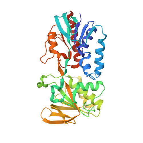
Structural and spectroscopic characterization of a HdrA-like subunit from Hyphomicrobium denitrificans.

Ernst, C.Kayastha, K.Koch, T.Venceslau, S.S.Pereira, I.A.C.Demmer, U.Ermler, U.Dahl, C.

(2021) FEBS J 288: 1664-1678

  • DOI: https://doi.org/10.1111/febs.15505
  • Primary Citation of Related Structures:  
    6TJR

  • PubMed Abstract: 

    Many bacteria and archaea employ a novel pathway of sulfur oxidation involving an enzyme complex that is related to the heterodisulfide reductase (Hdr or HdrABC) of methanogens. As a first step in the biochemical characterization of Hdr-like proteins from sulfur oxidizers (sHdr), we structurally analyzed the recombinant sHdrA protein from the Alphaproteobacterium Hyphomicrobium denitrificans at 1.4 Å resolution. The sHdrA core structure is similar to that of methanogenic HdrA (mHdrA) which binds the electron-bifurcating flavin adenine dinucleotide (FAD), the heart of the HdrABC-[NiFe]-hydrogenase catalyzed reaction. Each sHdrA homodimer carries two FADs and two [4Fe-4S] clusters being linked by electron conductivity. Redox titrations monitored by electron paramagnetic resonance and visible spectroscopy revealed a redox potential between -203 and -188 mV for the [4Fe-4S] center. The potentials for the FADH•/FADH - and FAD/FADH• pairs reside between -174 and -156 mV and between -81 and -19 mV, respectively. The resulting stable semiquinone FADH• species already detectable in the visible and electron paramagnetic resonance spectra of the as-isolated state of sHdrA is incompatible with basic principles of flavin-based electron bifurcation such that the sHdr complex does not apply this new mode of energy coupling. The inverted one-electron FAD redox potentials of sHdr and mHdr are clearly reflected in the different FAD-polypeptide interactions. According to this finding and the assumption that the sHdr complex forms an asymmetric HdrAA'B1C1B2C2 hexamer, we tentatively propose a mechanism that links protein-bound sulfane oxidation to sulfite on HdrB1 with NAD + reduction via lipoamide disulfide reduction on HdrB2. The FAD of HdrA thereby serves as an electron storage unit. DATABASE: Structural data are available in PDB database under the accession number 6TJR.


  • Organizational Affiliation

    Institut für Mikrobiologie & Biotechnologie, Rheinische Friedrich-Wilhelms-Universität Bonn, Bonn, Germany.


Macromolecules
Find similar proteins by:  (by identity cutoff)  |  3D Structure
Entity ID: 1
MoleculeChains Sequence LengthOrganismDetailsImage
Fumarate reductase/succinate dehydrogenase flavoprotein domain protein
A, B
349Hyphomicrobium denitrificansMutation(s): 0 
Gene Names: Hden_0691
UniProt
Find proteins for D8JT26 (Hyphomicrobium denitrificans (strain ATCC 51888 / DSM 1869 / NCIMB 11706 / TK 0415))
Explore D8JT26 
Go to UniProtKB:  D8JT26
Entity Groups  
Sequence Clusters30% Identity50% Identity70% Identity90% Identity95% Identity100% Identity
UniProt GroupD8JT26
Sequence Annotations
Expand
  • Reference Sequence
Small Molecules
Ligands 3 Unique
IDChains Name / Formula / InChI Key2D Diagram3D Interactions
FAD (Subject of Investigation/LOI)
Query on FAD

Download Ideal Coordinates CCD File 
D [auth A],
I [auth B]
FLAVIN-ADENINE DINUCLEOTIDE
C27 H33 N9 O15 P2
VWWQXMAJTJZDQX-UYBVJOGSSA-N
SF4 (Subject of Investigation/LOI)
Query on SF4

Download Ideal Coordinates CCD File 
C [auth A],
H [auth B]
IRON/SULFUR CLUSTER
Fe4 S4
LJBDFODJNLIPKO-UHFFFAOYSA-N
MES (Subject of Investigation/LOI)
Query on MES

Download Ideal Coordinates CCD File 
E [auth A],
F [auth A],
G [auth A],
J [auth B],
K [auth B]
2-(N-MORPHOLINO)-ETHANESULFONIC ACID
C6 H13 N O4 S
SXGZJKUKBWWHRA-UHFFFAOYSA-N
Experimental Data & Validation

Experimental Data

  • Method: X-RAY DIFFRACTION
  • Resolution: 1.43 Å
  • R-Value Free: 0.179 
  • R-Value Work: 0.163 
  • R-Value Observed: 0.164 
  • Space Group: P 65
Unit Cell:
Length ( Å )Angle ( ˚ )
a = 145.62α = 90
b = 145.62β = 90
c = 64.1γ = 120
Software Package:
Software NamePurpose
BUSTERrefinement
PDB_EXTRACTdata extraction
ARP/wARPmodel building
XSCALEdata scaling
SHARPphasing
XDSdata reduction

Structure Validation

View Full Validation Report


Ligand Structure Quality Assessment 


Entry History & Funding Information

Deposition Data


Funding OrganizationLocationGrant Number
German Research FoundationGermanyDa 351/8-1

Revision History  (Full details and data files)

  • Version 1.0: 2020-08-19
    Type: Initial release
  • Version 1.1: 2021-03-10
    Changes: Database references