6THY

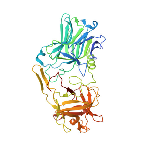
Botulinum neurotoxin A3 Hc domain in complex with GD1a


Experimental Data Snapshot

  • Method: X-RAY DIFFRACTION
  • Resolution: 1.75 Å
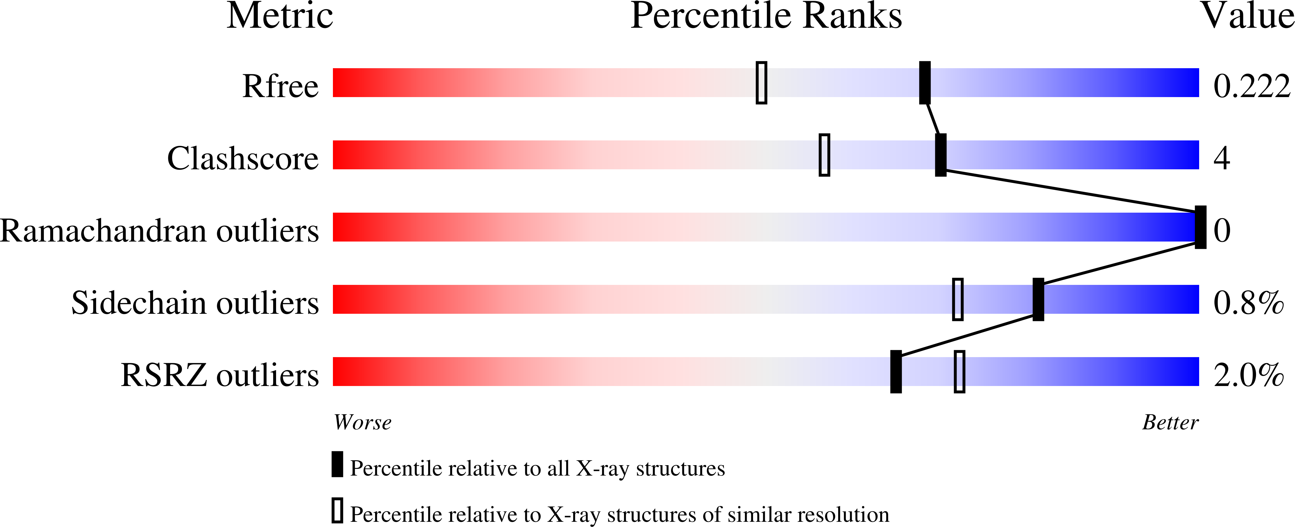
  • R-Value Free: 0.213 
  • R-Value Work: 0.178 

wwPDB Validation   3D Report Full Report


This is version 2.1 of the entry. See complete history


Literature

Crystal structure of botulinum neurotoxin subtype A3 cell binding domain in complex with GD1a co-receptor ganglioside.

Gregory, K.S.Liu, S.M.Acharya, K.R.

(2020) FEBS Open Bio 10: 298-305

  • DOI: https://doi.org/10.1002/2211-5463.12790
  • Primary Citation of Related Structures:  
    6THY

  • PubMed Abstract: 

    Botulinum neurotoxins (BoNTs) are one of the most toxic proteins known to humans. Their molecular structure is comprised of three essential domains-a cell binding domain (H C ), translocation domain and catalytic domain (light chain) . The H C domain facilitates the highly specific binding of BoNTs to the neuronal membrane via a dual-receptor complex involving a protein receptor and a ganglioside. Variation in activity/toxicity across subtypes of serotype A has been attributed to changes in protein and ganglioside interactions, and their implications are important in the design of novel BoNT-based therapeutics. Here, we present the structure of BoNT/A3 cell binding domain (H C /A3) in complex with the ganglioside GD1a at 1.75 Å resolution. The structure revealed that six residues interact with the three outermost monosaccharides of GD1a through several key hydrogen bonding interactions. A detailed comparison of structures of H C /A3 with H C /A1 revealed subtle conformational differences at the ganglioside binding site upon carbohydrate binding.


  • Organizational Affiliation

    Department of Biology and Biochemistry, Claverton Down, University of Bath, UK.


Macromolecules
Find similar proteins by:  (by identity cutoff)  |  3D Structure
Entity ID: 1
MoleculeChains Sequence LengthOrganismDetailsImage
BoNT/A3A [auth AAA]433Clostridium botulinumMutation(s): 0 
UniProt
Find proteins for Q3LRX9 (Clostridium botulinum)
Explore Q3LRX9 
Go to UniProtKB:  Q3LRX9
Entity Groups  
Sequence Clusters30% Identity50% Identity70% Identity90% Identity95% Identity100% Identity
UniProt GroupQ3LRX9
Sequence Annotations
Expand
  • Reference Sequence
Oligosaccharides

Help

Entity ID: 2
MoleculeChains Length2D Diagram Glycosylation3D Interactions
N-acetyl-alpha-neuraminic acid-(2-3)-beta-D-galactopyranose-(1-3)-2-acetamido-2-deoxy-beta-D-galactopyranose-(1-4)-beta-D-galactopyranose-(1-4)-beta-D-glucopyranoseB [auth A]5N/A
Glycosylation Resources
GlyTouCan:  G03277YI
GlyCosmos:  G03277YI
GlyGen:  G03277YI
Small Molecules
Ligands 4 Unique
IDChains Name / Formula / InChI Key2D Diagram3D Interactions
P6G
Query on P6G

Download Ideal Coordinates CCD File 
C [auth AAA]HEXAETHYLENE GLYCOL
C12 H26 O7
IIRDTKBZINWQAW-UHFFFAOYSA-N
PEG
Query on PEG

Download Ideal Coordinates CCD File 
G [auth AAA],
H [auth AAA]
DI(HYDROXYETHYL)ETHER
C4 H10 O3
MTHSVFCYNBDYFN-UHFFFAOYSA-N
EDO
Query on EDO

Download Ideal Coordinates CCD File 
D [auth AAA],
F [auth AAA]
1,2-ETHANEDIOL
C2 H6 O2
LYCAIKOWRPUZTN-UHFFFAOYSA-N
ACT
Query on ACT

Download Ideal Coordinates CCD File 
E [auth AAA]ACETATE ION
C2 H3 O2
QTBSBXVTEAMEQO-UHFFFAOYSA-M
Experimental Data & Validation

Experimental Data

  • Method: X-RAY DIFFRACTION
  • Resolution: 1.75 Å
  • R-Value Free: 0.213 
  • R-Value Work: 0.178 
  • Space Group: P 21 21 21
Unit Cell:
Length ( Å )Angle ( ˚ )
a = 45.228α = 90
b = 73.129β = 90
c = 140.18γ = 90
Software Package:
Software NamePurpose
REFMACrefinement
DIALSdata reduction
Aimlessdata scaling
PHASERphasing

Structure Validation

View Full Validation Report


Entry History 

Deposition Data

Revision History  (Full details and data files)

  • Version 1.0: 2020-01-29
    Type: Initial release
  • Version 1.1: 2020-03-11
    Changes: Data collection, Database references
  • Version 2.0: 2020-07-29
    Type: Remediation
    Reason: Carbohydrate remediation
    Changes: Atomic model, Author supporting evidence, Data collection, Derived calculations, Structure summary
  • Version 2.1: 2024-01-24
    Changes: Data collection, Database references, Derived calculations, Refinement description, Structure summary