


Crystal structure of the CheY in presence of magnesium
Camara-Artigas, A., Salinas-Garcia, M.C., Alba-Elena, D.To be published.
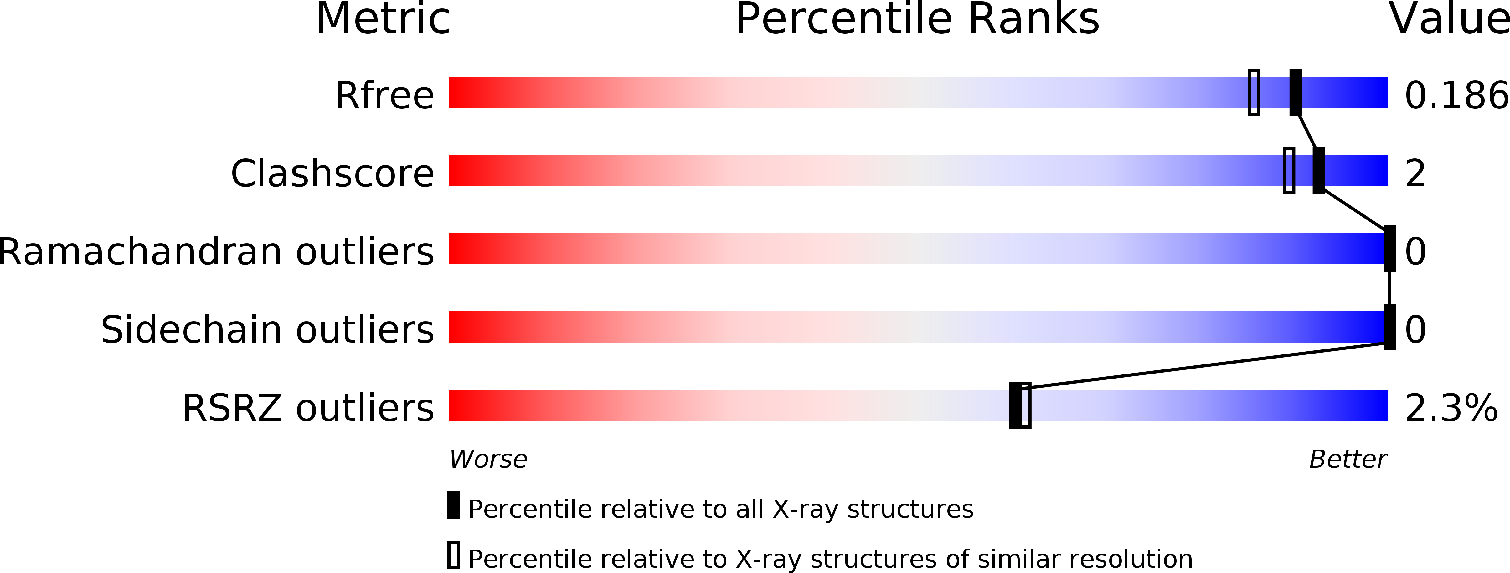
Experimental Data Snapshot
wwPDB Validation 3D Report Full Report
Entity ID: 1 | |||||
|---|---|---|---|---|---|
| Molecule | Chains | Sequence Length | Organism | Details | Image |
| Chemotaxis protein CheY | 155 | Escherichia coli 5-366-08_S1_C3 | Mutation(s): 0 Gene Names: cheY, AB67_2120 | 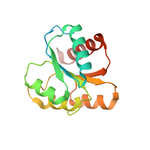 | |
UniProt | |||||
Find proteins for P0AE67 (Escherichia coli (strain K12)) Explore P0AE67 Go to UniProtKB: P0AE67 | |||||
Entity Groups | |||||
| Sequence Clusters | 30% Identity50% Identity70% Identity90% Identity95% Identity100% Identity | ||||
| UniProt Group | P0AE67 | ||||
Sequence AnnotationsExpand | |||||
| |||||
| Ligands 1 Unique | |||||
|---|---|---|---|---|---|
| ID | Chains | Name / Formula / InChI Key | 2D Diagram | 3D Interactions | |
| MG Query on MG | B [auth A] | MAGNESIUM ION Mg JLVVSXFLKOJNIY-UHFFFAOYSA-N |  | ||
| Length ( Å ) | Angle ( ˚ ) |
|---|---|
| a = 45.399 | α = 90 |
| b = 46.924 | β = 90 |
| c = 53.198 | γ = 90 |
| Software Name | Purpose |
|---|---|
| PHENIX | refinement |
| XDS | data reduction |
| Aimless | data scaling |
| PHASER | phasing |
| PDB_EXTRACT | data extraction |
| Funding Organization | Location | Grant Number |
|---|---|---|
| Spanish Ministry of Economy and Competitiveness | Spain | BIO2016-78020-R |
RCSB PDB (citation) is hosted by
RCSB PDB is a member of the

