6SYF

Human Ubc9 with covalent isopeptide ligand


Experimental Data Snapshot

  • Method: X-RAY DIFFRACTION
  • Resolution: 1.90 Å
  • R-Value Free: 0.263 
  • R-Value Work: 0.213 
  • R-Value Observed: 0.215 

wwPDB Validation   3D Report Full Report


This is version 1.4 of the entry. See complete history


Literature

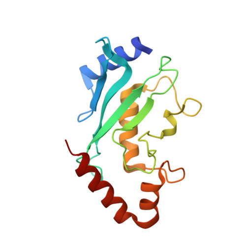
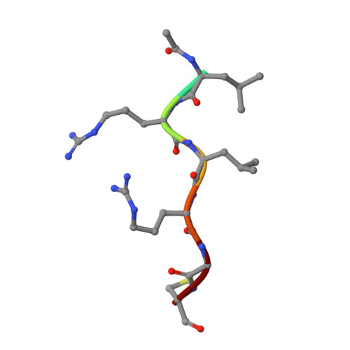
Lysine acylation using conjugating enzymes for site-specific modification and ubiquitination of recombinant proteins.

Hofmann, R.Akimoto, G.Wucherpfennig, T.G.Zeymer, C.Bode, J.W.

(2020) Nat Chem 12: 1008-1015

  • DOI: https://doi.org/10.1038/s41557-020-0528-y
  • Primary Citation of Related Structures:  
    6SYF

  • PubMed Abstract: 

    Enzymes are powerful tools for protein labelling due to their specificity and mild reaction conditions. Many protocols, however, are restricted to modifications at protein termini, rely on non-peptidic metabolites or require large recognition domains. Here we report a chemoenzymatic method, which we call lysine acylation using conjugating enzymes (LACE), to site-specifically modify folded proteins at internal lysine residues. LACE relies on a minimal genetically encoded tag (four residues) recognized by the E2 small ubiquitin-like modifier-conjugating enzyme Ubc9, and peptide or protein thioesters. Together, this approach obviates the need for E1 and E3 enzymes, enabling isopeptide formation with just Ubc9 in a programmable manner. We demonstrate the utility of LACE by the site-specific attachment of biochemical probes, one-pot dual-labelling in combination with sortase, and the conjugation of wild-type ubiquitin and ISG15 to recombinant proteins.


  • Organizational Affiliation

    Laboratorium für Organische Chemie, Department of Chemistry and Applied Biosciences, ETH Zürich, Zürich, Switzerland.


Macromolecules
Find similar proteins by:  (by identity cutoff)  |  3D Structure
Entity ID: 1
MoleculeChains Sequence LengthOrganismDetailsImage
SUMO-conjugating enzyme UBC9
A, B, C, D
158Homo sapiensMutation(s): 4 
Gene Names: UBE2IUBC9UBCE9
EC: 2.3.2
UniProt & NIH Common Fund Data Resources
Find proteins for P63279 (Homo sapiens)
Explore P63279 
Go to UniProtKB:  P63279
PHAROS:  P63279
GTEx:  ENSG00000103275 
Entity Groups  
Sequence Clusters30% Identity50% Identity70% Identity90% Identity95% Identity100% Identity
UniProt GroupP63279
Sequence Annotations
Expand
  • Reference Sequence

Find similar proteins by:  Sequence   |   3D Structure  

Entity ID: 2
MoleculeChains Sequence LengthOrganismDetailsImage
ACE-LEU-ARG-LEU-ARG-GLY-CYS
E, G, I, K
7Homo sapiensMutation(s): 0 
Sequence Annotations
Expand
  • Reference Sequence

Find similar proteins by:  Sequence   |   3D Structure  

Entity ID: 3
MoleculeChains Sequence LengthOrganismDetailsImage
ACE-ILE-LYS-GLN-GLU
F, H, J, L
5Homo sapiensMutation(s): 0 
Sequence Annotations
Expand
  • Reference Sequence
Experimental Data & Validation

Experimental Data

  • Method: X-RAY DIFFRACTION
  • Resolution: 1.90 Å
  • R-Value Free: 0.263 
  • R-Value Work: 0.213 
  • R-Value Observed: 0.215 
  • Space Group: P 1 21 1
Unit Cell:
Length ( Å )Angle ( ˚ )
a = 94.7α = 90
b = 38.7β = 118.9
c = 97.8γ = 90
Software Package:
Software NamePurpose
XDSdata reduction
XSCALEdata scaling
PHASERphasing
BUSTERrefinement
PDB_EXTRACTdata extraction

Structure Validation

View Full Validation Report


Entry History 

Deposition Data

Revision History  (Full details and data files)

  • Version 1.0: 2020-08-12
    Type: Initial release
  • Version 1.1: 2020-09-16
    Changes: Database references
  • Version 1.2: 2020-09-23
    Changes: Database references
  • Version 1.3: 2020-11-04
    Changes: Database references
  • Version 1.4: 2024-01-24
    Changes: Data collection, Database references, Refinement description