6SWT

Affimer9 co-crystalised with the CH domains of alpha actinin 2.


Experimental Data Snapshot

  • Method: X-RAY DIFFRACTION
  • Resolution: 1.20 Å
  • R-Value Free: 0.164 
  • R-Value Work: 0.149 
  • R-Value Observed: 0.150 

wwPDB Validation   3D Report Full Report


This is version 1.3 of the entry. See complete history


Literature

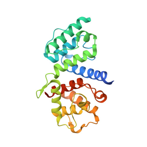
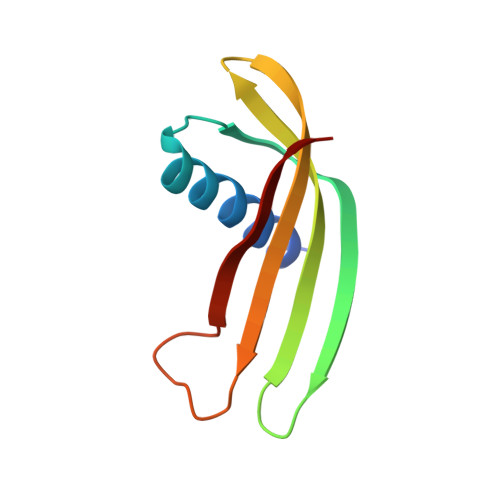
Nanoscale Pattern Extraction from Relative Positions of Sparse 3D Localizations.

Curd, A.P.Leng, J.Hughes, R.E.Cleasby, A.J.Rogers, B.Trinh, C.H.Baird, M.A.Takagi, Y.Tiede, C.Sieben, C.Manley, S.Schlichthaerle, T.Jungmann, R.Ries, J.Shroff, H.Peckham, M.

(2021) Nano Lett 21: 1213-1220

  • DOI: https://doi.org/10.1021/acs.nanolett.0c03332
  • Primary Citation of Related Structures:  
    6SWT

  • PubMed Abstract: 

    Inferring the organization of fluorescently labeled nanosized structures from single molecule localization microscopy (SMLM) data, typically obscured by stochastic noise and background, remains challenging. To overcome this, we developed a method to extract high-resolution ordered features from SMLM data that requires only a low fraction of targets to be localized with high precision. First, experimentally measured localizations are analyzed to produce relative position distributions (RPDs). Next, model RPDs are constructed using hypotheses of how the molecule is organized. Finally, a statistical comparison is used to select the most likely model. This approach allows pattern recognition at sub-1% detection efficiencies for target molecules, in large and heterogeneous samples and in 2D and 3D data sets. As a proof-of-concept, we infer ultrastructure of Nup107 within the nuclear pore, DNA origami structures, and α-actinin-2 within the cardiomyocyte Z-disc and assess the quality of images of centrioles to improve the averaged single-particle reconstruction.


  • Organizational Affiliation

    School of Molecular and Cellular Biology, University of Leeds, Leeds LS2 9JT, United Kingdom.


Macromolecules
Find similar proteins by:  (by identity cutoff)  |  3D Structure
Entity ID: 1
MoleculeChains Sequence LengthOrganismDetailsImage
Alpha-actinin-2254Homo sapiensMutation(s): 0 
Gene Names: ACTN2
UniProt & NIH Common Fund Data Resources
Find proteins for P35609 (Homo sapiens)
Explore P35609 
Go to UniProtKB:  P35609
PHAROS:  P35609
GTEx:  ENSG00000077522 
Entity Groups  
Sequence Clusters30% Identity50% Identity70% Identity90% Identity95% Identity100% Identity
UniProt GroupP35609
Sequence Annotations
Expand
  • Reference Sequence
Find similar proteins by:  (by identity cutoff)  |  3D Structure
Entity ID: 2
MoleculeChains Sequence LengthOrganismDetailsImage
Affimer 991synthetic constructMutation(s): 0 
Entity Groups  
Sequence Clusters30% Identity50% Identity70% Identity90% Identity95% Identity100% Identity
Sequence Annotations
Expand
  • Reference Sequence
Experimental Data & Validation

Experimental Data

  • Method: X-RAY DIFFRACTION
  • Resolution: 1.20 Å
  • R-Value Free: 0.164 
  • R-Value Work: 0.149 
  • R-Value Observed: 0.150 
  • Space Group: P 21 21 21
Unit Cell:
Length ( Å )Angle ( ˚ )
a = 46.192α = 90
b = 48.519β = 90
c = 146.993γ = 90
Software Package:
Software NamePurpose
DIALSdata reduction
Aimlessdata scaling
PHASERphasing
REFMACrefinement

Structure Validation

View Full Validation Report


Entry History & Funding Information

Deposition Data


Funding OrganizationLocationGrant Number
Medical Research Council (United Kingdom)United KingdomMR/K015613/1
Biotechnology and Biological Sciences Research CouncilUnited KingdomBB/M011151/1

Revision History  (Full details and data files)

  • Version 1.0: 2020-10-07
    Type: Initial release
  • Version 1.1: 2020-12-09
    Changes: Database references
  • Version 1.2: 2021-02-17
    Changes: Database references
  • Version 1.3: 2024-01-24
    Changes: Data collection, Database references, Refinement description