6SVA

Multicrystal structure of equine Haemoglobin at room temperature using a multilayer monochromator.


Experimental Data Snapshot

  • Method: X-RAY DIFFRACTION
  • Resolution: 1.92 Å
  • R-Value Free: 0.173 
  • R-Value Work: 0.141 
  • R-Value Observed: 0.142 

wwPDB Validation   3D Report Full Report


Ligand Structure Quality Assessment 


This is version 1.2 of the entry. See complete history


Literature

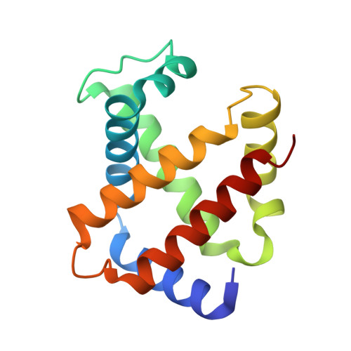
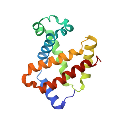
Protein-to-structure pipeline for ambient-temperature crystallography at VMXi

Mikolajek, H.Sanchez-Weatherby, J.Sandy, J.Gildea, R.G.Campeotto, I.Cheruvara, H.Clarke, J.D.Foster, T.Fujii, S.Paulsen, I.T.Shah, B.S.Hough, M.A.

(2023) IUCrJ 


Macromolecules
Find similar proteins by:  (by identity cutoff)  |  3D Structure
Entity ID: 1
MoleculeChains Sequence LengthOrganismDetailsImage
Hemoglobin subunit alpha140Equus caballusMutation(s): 0 
UniProt
Find proteins for P01958 (Equus caballus)
Explore P01958 
Go to UniProtKB:  P01958
Entity Groups  
Sequence Clusters30% Identity50% Identity70% Identity90% Identity95% Identity100% Identity
UniProt GroupP01958
Sequence Annotations
Expand
  • Reference Sequence
Find similar proteins by:  (by identity cutoff)  |  3D Structure
Entity ID: 2
MoleculeChains Sequence LengthOrganismDetailsImage
Hemoglobin subunit beta146Equus caballusMutation(s): 0 
UniProt
Find proteins for P02062 (Equus caballus)
Explore P02062 
Go to UniProtKB:  P02062
Entity Groups  
Sequence Clusters30% Identity50% Identity70% Identity90% Identity95% Identity100% Identity
UniProt GroupP02062
Sequence Annotations
Expand
  • Reference Sequence
Small Molecules
Ligands 1 Unique
IDChains Name / Formula / InChI Key2D Diagram3D Interactions
HEM
Query on HEM

Download Ideal Coordinates CCD File 
C [auth A],
D [auth B]
PROTOPORPHYRIN IX CONTAINING FE
C34 H32 Fe N4 O4
KABFMIBPWCXCRK-RGGAHWMASA-L
Experimental Data & Validation

Experimental Data

  • Method: X-RAY DIFFRACTION
  • Resolution: 1.92 Å
  • R-Value Free: 0.173 
  • R-Value Work: 0.141 
  • R-Value Observed: 0.142 
  • Space Group: C 1 2 1
Unit Cell:
Length ( Å )Angle ( ˚ )
a = 108.515α = 90
b = 62.992β = 111.051
c = 54.591γ = 90
Software Package:
Software NamePurpose
PHENIXrefinement
PHENIXrefinement
DIALSdata reduction
xia2data scaling
PHASERphasing

Structure Validation

View Full Validation Report


Ligand Structure Quality Assessment 


Entry History 

Deposition Data

Revision History  (Full details and data files)

  • Version 1.0: 2019-11-20
    Type: Initial release
  • Version 1.1: 2023-05-17
    Changes: Database references, Derived calculations
  • Version 1.2: 2024-02-07
    Changes: Data collection, Refinement description