Structure of High-Risk Papillomavirus 31 E6 Oncogenic Protein and Characterization of E6/E6AP/p53 Complex Formation.
Conrady, M.C., Suarez, I., Gogl, G., Frecot, D.I., Bonhoure, A., Kostmann, C., Cousido-Siah, A., Mitschler, A., Lim, J., Masson, M., Iftner, T., Stubenrauch, F., Trave, G., Simon, C.(2020) J Virol 95
- PubMed: 33115863
- DOI: https://doi.org/10.1128/JVI.00730-20
- Primary Citation of Related Structures:
6SLM - PubMed Abstract:
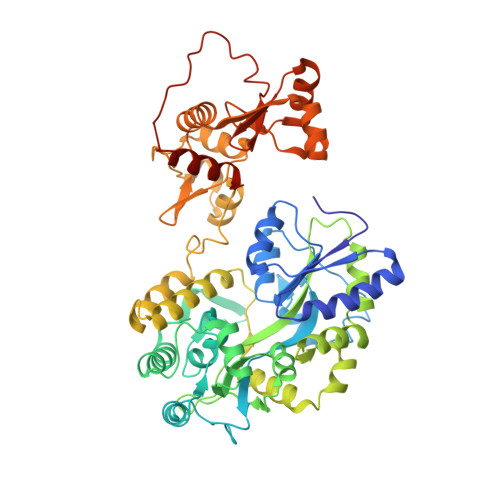
The degradation of p53 is a hallmark of high-risk human papillomaviruses (HPVs) of the alpha genus and HPV-related carcinogenicity. The oncoprotein E6 forms a ternary complex with the E3 ubiquitin ligase E6-associated protein (E6AP) and tumor suppressor protein p53 targeting p53 for ubiquitination. The extent of p53 degradation by different E6 proteins varies greatly, even for the closely related HPV16 and HPV31. HPV16 E6 and HPV31 E6 display high sequence identity (∼67%). We report here, for the first time, the structure of HPV31 E6 bound to the LxxLL motif of E6AP. HPV16 E6 and HPV31 E6 are structurally very similar, in agreement with the high sequence conservation. Both E6 proteins bind E6AP and degrade p53. However, the binding affinities of 31 E6 to the LxxLL motif of E6AP and p53, respectively, are reduced 2-fold and 5.4-fold compared to 16 E6. The affinity of E6-E6AP-p53 ternary complex formation parallels the efficacy of the subsequent reaction, namely, degradation of p53. Therefore, closely related E6 proteins addressing the same cellular targets may still diverge in their binding efficiencies, possibly explaining their different phenotypic or pathological impacts. IMPORTANCE Variations of carcinogenicity of human papillomaviruses are related to variations of the E6 and E7 interactome. While different HPV species and genera are known to target distinct host proteins, the fine differences between E6 and E7 of closely related HPVs, supposed to target the same cellular protein pools, remain to be addressed. We compare the oncogenic E6 proteins of the closely related high-risk HPV31 and HPV16 with regard to their structure and their efficiency of ternary complex formation with their cellular targets p53 and E6AP, which results in p53 degradation. We solved the crystal structure of 31 E6 bound to the E6AP LxxLL motif. HPV16 E6 and 31 E6 structures are highly similar, but a few sequence variations lead to different protein contacts within the ternary complex and, as quantified here, an overall lower binding affinity of 31 E6 than 16 E6. These results align with the observed lower p53 degradation potential of 31 E6.
Organizational Affiliation:
Institute of Medical Virology, Medical Faculty, Eberhard Karls University, Tuebingen, Germany.