6S0C

Crystal structure of methionine gamma-lyase from Citrobacter freundii modified by dimethylthiosulfinate


Experimental Data Snapshot

  • Method: X-RAY DIFFRACTION
  • Resolution: 1.46 Å
  • R-Value Free: 0.172 
  • R-Value Work: 0.137 
  • R-Value Observed: 0.138 

wwPDB Validation   3D Report Full Report


Ligand Structure Quality Assessment 


This is version 1.1 of the entry. See complete history


Literature

Sulfoxides of sulfur-containing amino acids are suicide substrates of Citrobacter freundii methionine gamma-lyase. Structural bases of the enzyme inactivation.

Revtovich, S.Morozova, E.Kulikova, V.Koval, V.Anufrieva, N.Nikulin, A.Demidkina, T.

(2020) Biochimie 168: 190-197

  • DOI: https://doi.org/10.1016/j.biochi.2019.11.004
  • Primary Citation of Related Structures:  
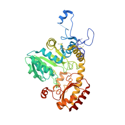
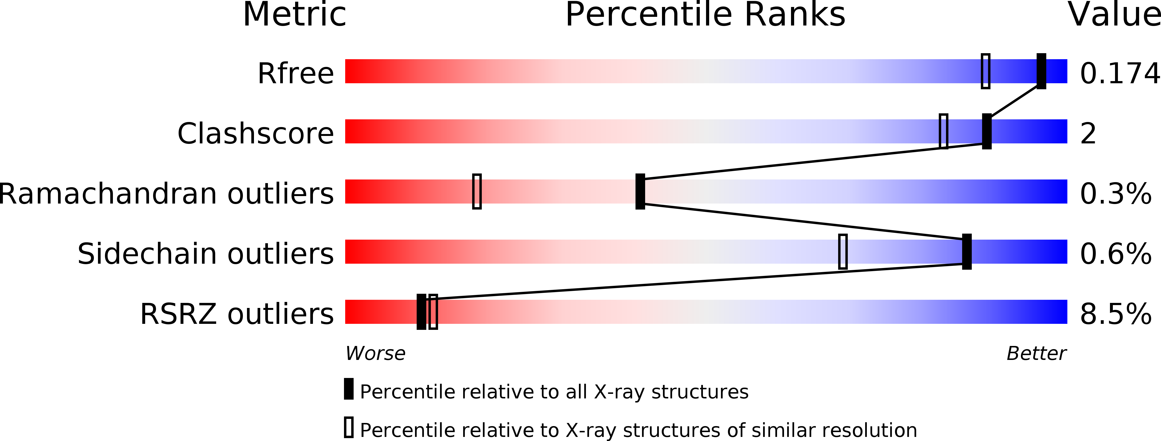
    5K30, 6S0C

  • PubMed Abstract: 

    Interactions of Citrobacter freundii methionine γ-lyase (MGL) with sulfoxides of typical substrates were investigated. It was found that sulfoxides are suicide substrates of the enzyme. The products of the β- and γ-elimination reactions of sulfoxides, thiosulfinates, oxidize three cysteine residues of the enzyme. Three-dimensional structures of MGL inactivated by dimethyl thiosulfinate and diethyl thiosulfinate were determined at 1.46 Å and 1.59 Å resolution. Analysis of the structures identified SH groups oxidized by thiosulfinates and revealed the structural bases of MGL inactivation. The extent of inactivation of MGL in the catalysis of the β-elimination reaction depends on the length of the «tail» at oxidized Cys115. Oxidation of Cys115 results in MGL incapable to catalyze the stage of methyl mercaptan elimination of the physiological reaction.


  • Organizational Affiliation

    Engelhardt Institute of Molecular Biology of the Russian Academy of Sciences, Moscow, Russia. Electronic address: svetla21@mail.ru.


Macromolecules
Find similar proteins by:  (by identity cutoff)  |  3D Structure
Entity ID: 1
MoleculeChains Sequence LengthOrganismDetailsImage
Cystathionine gamma-synthase398Citrobacter freundiiMutation(s): 0 
Gene Names: 
EC: 4.4.1.11
UniProt
Find proteins for A0A0A5P8W7 (Citrobacter freundii)
Explore A0A0A5P8W7 
Go to UniProtKB:  A0A0A5P8W7
Entity Groups  
Sequence Clusters30% Identity50% Identity70% Identity90% Identity95% Identity100% Identity
UniProt GroupA0A0A5P8W7
Sequence Annotations
Expand
  • Reference Sequence
Small Molecules
Ligands 2 Unique
IDChains Name / Formula / InChI Key2D Diagram3D Interactions
PLP
Query on PLP

Download Ideal Coordinates CCD File 
C [auth A]PYRIDOXAL-5'-PHOSPHATE
C8 H10 N O6 P
NGVDGCNFYWLIFO-UHFFFAOYSA-N
PGE
Query on PGE

Download Ideal Coordinates CCD File 
B [auth A]TRIETHYLENE GLYCOL
C6 H14 O4
ZIBGPFATKBEMQZ-UHFFFAOYSA-N
Modified Residues  1 Unique
IDChains TypeFormula2D DiagramParent
SCH
Query on SCH
A
L-PEPTIDE LINKINGC4 H9 N O2 S2CYS
Experimental Data & Validation

Experimental Data

  • Method: X-RAY DIFFRACTION
  • Resolution: 1.46 Å
  • R-Value Free: 0.172 
  • R-Value Work: 0.137 
  • R-Value Observed: 0.138 
  • Space Group: I 2 2 2
Unit Cell:
Length ( Å )Angle ( ˚ )
a = 56.784α = 90
b = 123.255β = 90
c = 128.328γ = 90
Software Package:
Software NamePurpose
PHENIXrefinement
XDSdata reduction
Aimlessdata scaling
PHASERphasing

Structure Validation

View Full Validation Report


Ligand Structure Quality Assessment 


Entry History & Funding Information

Deposition Data


Funding OrganizationLocationGrant Number
Russian Science FoundationRussian Federation15-14-00009

Revision History  (Full details and data files)

  • Version 1.0: 2020-04-22
    Type: Initial release
  • Version 1.1: 2024-01-24
    Changes: Data collection, Database references, Refinement description