


Crystal structure of Escherichia coli Glyoxalase II
Skorupskaite, A., McDonough, M.A., Brem, J., Schofield, C.J.To be published.
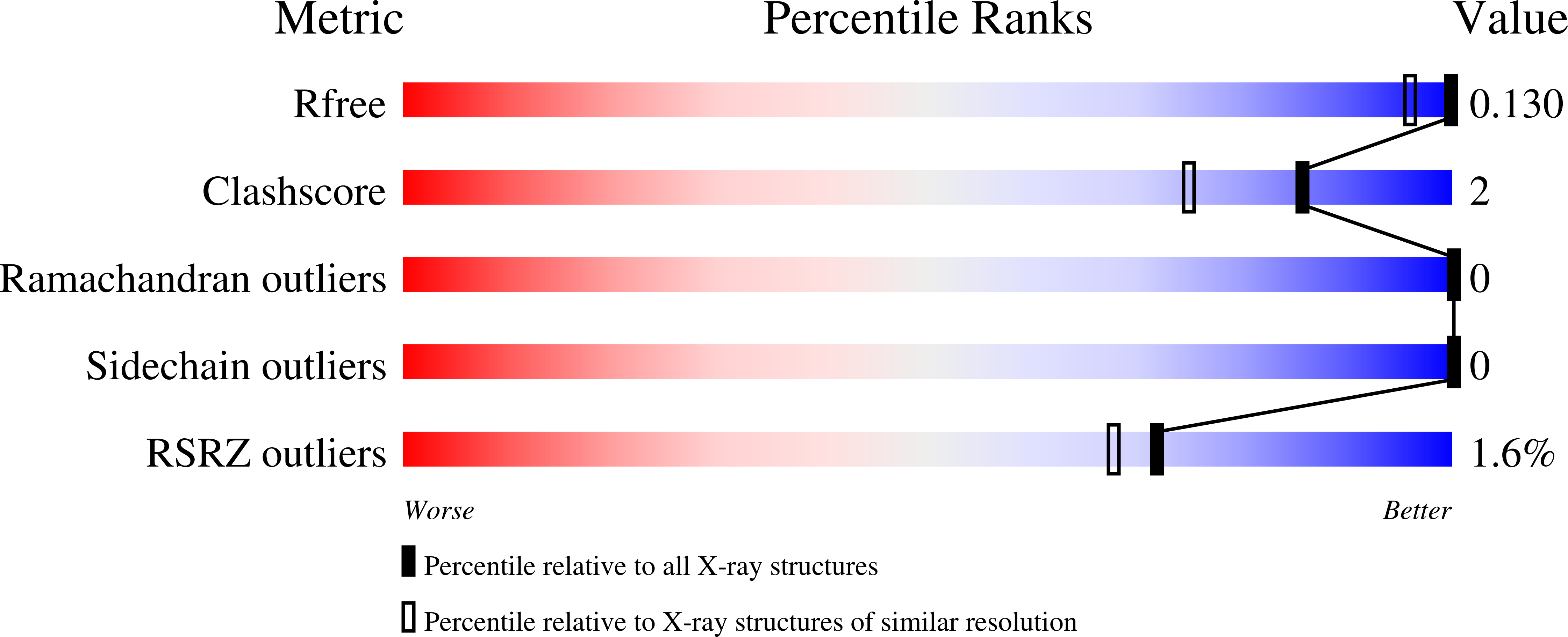
Experimental Data Snapshot
Entity ID: 1 | |||||
|---|---|---|---|---|---|
| Molecule | Chains | Sequence Length | Organism | Details | Image |
| Hydroxyacylglutathione hydrolase GloB | 251 | Escherichia coli K-12 | Mutation(s): 0 Gene Names: gloB, yafR, b0212, JW0202 EC: 3.1.2.6 | 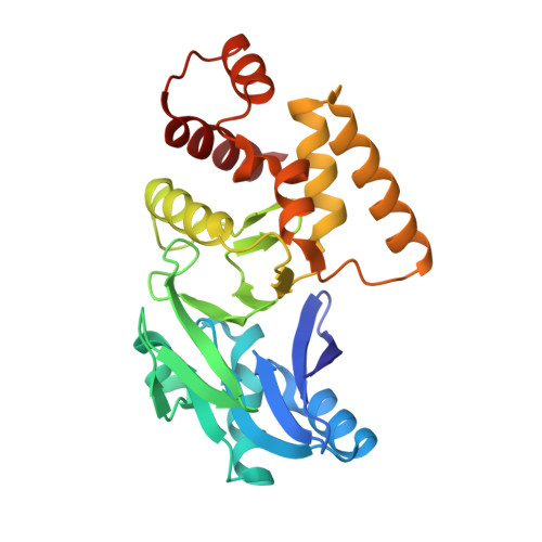 | |
UniProt | |||||
Find proteins for P0AC84 (Escherichia coli (strain K12)) Explore P0AC84 Go to UniProtKB: P0AC84 | |||||
Entity Groups | |||||
| Sequence Clusters | 30% Identity50% Identity70% Identity90% Identity95% Identity100% Identity | ||||
| UniProt Group | P0AC84 | ||||
Sequence AnnotationsExpand | |||||
| |||||
| Ligands 4 Unique | |||||
|---|---|---|---|---|---|
| ID | Chains | Name / Formula / InChI Key | 2D Diagram | 3D Interactions | |
| CXS Query on CXS | F [auth A] | 3-CYCLOHEXYL-1-PROPYLSULFONIC ACID C9 H19 N O3 S PJWWRFATQTVXHA-UHFFFAOYSA-N |  | ||
| GOL Query on GOL | B [auth A] | GLYCEROL C3 H8 O3 PEDCQBHIVMGVHV-UHFFFAOYSA-N |  | ||
| ZN Query on ZN | C [auth A], D [auth A], E [auth A], I [auth A] | ZINC ION Zn PTFCDOFLOPIGGS-UHFFFAOYSA-N |  | ||
| CL Query on CL | G [auth A], H [auth A] | CHLORIDE ION Cl VEXZGXHMUGYJMC-UHFFFAOYSA-M |  | ||
| Length ( Å ) | Angle ( ˚ ) |
|---|---|
| a = 59.382 | α = 90 |
| b = 63.383 | β = 90 |
| c = 72.726 | γ = 90 |
| Software Name | Purpose |
|---|---|
| PHENIX | refinement |
| XDS | data reduction |
| Aimless | data scaling |
| PHASER | phasing |
| PDB_EXTRACT | data extraction |
| Funding Organization | Location | Grant Number |
|---|---|---|
| Biotechnology and Biological Sciences Research Council | United Kingdom | BB/M011224/1 |
RCSB PDB (citation) is hosted by
RCSB PDB is a member of the

