


CRYSTAL STRUCTURE OF the VhH-domain of anti-IL-17A antibody netakimab
Kostareva, O.S., Kolyadenko, I.A., Ulitin, A.B., Ekimova, V.M., Evdokimov, S.R., Garber, M.B., Tishchenko, T.V., Gabdulkhakov, A.G.To be published.
Experimental Data Snapshot
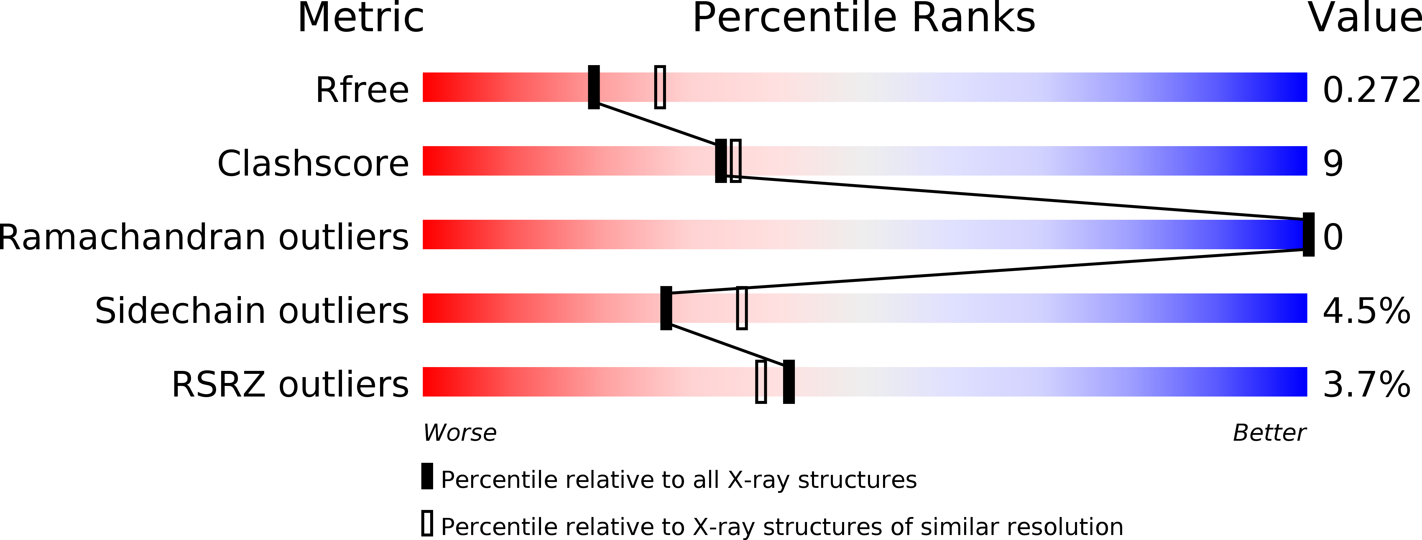
wwPDB Validation 3D Report Full Report
Entity ID: 1 | |||||
|---|---|---|---|---|---|
| Molecule | Chains | Sequence Length | Organism | Details | Image |
| VHH domain of netakimab | A [auth H], B [auth I], C [auth J], D [auth K] | 123 | Lama glama | Mutation(s): 0 | 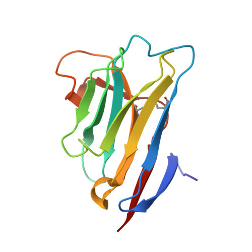 |
Entity Groups | |||||
| Sequence Clusters | 30% Identity50% Identity70% Identity90% Identity95% Identity100% Identity | ||||
Sequence AnnotationsExpand | |||||
| |||||
| Length ( Å ) | Angle ( ˚ ) |
|---|---|
| a = 54.872 | α = 90 |
| b = 72.499 | β = 90 |
| c = 129.385 | γ = 90 |
| Software Name | Purpose |
|---|---|
| PHENIX | refinement |
| XDS | data reduction |
| XSCALE | data scaling |
| PHASER | phasing |
| Funding Organization | Location | Grant Number |
|---|---|---|
| Russian Science Foundation | Russian Federation | 17-74-10156 |
RCSB PDB (citation) is hosted by
RCSB PDB is a member of the

