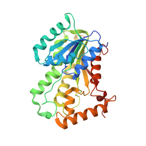
Discovery of New and Potent InhA Inhibitors as Antituberculosis Agents: Structure-Based Virtual Screening Validated by Biological Assays and X-ray Crystallography.
Kamsri, P., Hanwarinroj, C., Phusi, N., Pornprom, T., Chayajarus, K., Punkvang, A., Suttipanta, N., Srimanote, P., Suttisintong, K., Songsiriritthigul, C., Saparpakorn, P., Hannongbua, S., Rattanabunyong, S., Seetaha, S., Choowongkomon, K., Sureram, S., Kittakoop, P., Hongmanee, P., Santanirand, P., Chen, Z., Zhu, W., Blood, R.A., Takebayashi, Y., Hinchliffe, P., Mulholland, A.J., Spencer, J., Pungpo, P.(2020) J Chem Inf Model 60: 226-234
- PubMed: 31820972
- DOI: https://doi.org/10.1021/acs.jcim.9b00918
- Primary Citation of Related Structures:
6R9W - PubMed Abstract:
The enoyl-acyl carrier protein reductase InhA of Mycobacterium tuberculosis is an attractive, validated target for antituberculosis drug development. Moreover, direct inhibitors of InhA remain effective against InhA variants with mutations associated with isoniazid resistance, offering the potential for activity against MDR isolates. Here, structure-based virtual screening supported by biological assays was applied to identify novel InhA inhibitors as potential antituberculosis agents. High-speed Glide SP docking was initially performed against two conformations of InhA differing in the orientation of the active site Tyr158. The resulting hits were filtered for drug-likeness based on Lipinski's rule and avoidance of PAINS-like properties and finally subjected to Glide XP docking to improve accuracy. Sixteen compounds were identified and selected for in vitro biological assays, of which two (compounds 1 and 7 ) showed MIC of 12.5 and 25 μg/mL against M. tuberculosis H37Rv, respectively. Inhibition assays against purified recombinant InhA determined IC 50 values for these compounds of 0.38 and 0.22 μM, respectively. A crystal structure of the most potent compound, compound 7 , bound to InhA revealed the inhibitor to occupy a hydrophobic pocket implicated in binding the aliphatic portions of InhA substrates but distant from the NADH cofactor, i.e., in a site distinct from those occupied by the great majority of known InhA inhibitors. This compound provides an attractive starting template for ligand optimization aimed at discovery of new and effective compounds against M. tuberculosis that act by targeting InhA.
Organizational Affiliation:
Division of Chemistry, Faculty of Science , Nakhon Phanom University , 48000 Nakhon Phanom , Thailand.


