6QTI

Structure of ovine transhydrogenase in the presence of NADP+ in a "double face-down" conformation


Experimental Data Snapshot

  • Method: ELECTRON MICROSCOPY
  • Resolution: 2.90 Å
  • Aggregation State: PARTICLE 
  • Reconstruction Method: SINGLE PARTICLE 

wwPDB Validation   3D Report Full Report


This is version 1.3 of the entry. See complete history


Literature

Structure and mechanism of mitochondrial proton-translocating transhydrogenase.

Kampjut, D.Sazanov, L.A.

(2019) Nature 573: 291-295

  • DOI: https://doi.org/10.1038/s41586-019-1519-2
  • Primary Citation of Related Structures:  
    6QTI, 6QUE, 6S59

  • PubMed Abstract: 

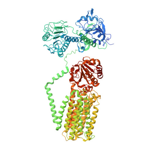
    Proton-translocating transhydrogenase (also known as nicotinamide nucleotide transhydrogenase (NNT)) is found in the plasma membranes of bacteria and the inner mitochondrial membranes of eukaryotes. NNT catalyses the transfer of a hydride between NADH and NADP + , coupled to the translocation of one proton across the membrane. Its main physiological function is the generation of NADPH, which is a substrate in anabolic reactions and a regulator of oxidative status; however, NNT may also fine-tune the Krebs cycle 1,2 . NNT deficiency causes familial glucocorticoid deficiency in humans and metabolic abnormalities in mice, similar to those observed in type II diabetes 3,4 . The catalytic mechanism of NNT has been proposed to involve a rotation of around 180° of the entire NADP(H)-binding domain that alternately participates in hydride transfer and proton-channel gating. However, owing to the lack of high-resolution structures of intact NNT, the details of this process remain unclear 5,6 . Here we present the cryo-electron microscopy structure of intact mammalian NNT in different conformational states. We show how the NADP(H)-binding domain opens the proton channel to the opposite sides of the membrane, and we provide structures of these two states. We also describe the catalytically important interfaces and linkers between the membrane and the soluble domains and their roles in nucleotide exchange. These structures enable us to propose a revised mechanism for a coupling process in NNT that is consistent with a large body of previous biochemical work. Our results are relevant to the development of currently unavailable NNT inhibitors, which may have therapeutic potential in ischaemia reperfusion injury, metabolic syndrome and some cancers 7-9 .


  • Organizational Affiliation

    Institute of Science and Technology Austria, Klosterneuburg, Austria.


Macromolecules
Find similar proteins by:  (by identity cutoff)  |  3D Structure
Entity ID: 1
MoleculeChains Sequence LengthOrganismDetailsImage
Nicotinamide nucleotide transhydrogenase
A, B
1,086Ovis ariesMutation(s): 0 
Membrane Entity: Yes 
UniProt
Find proteins for W5PFI3 (Ovis aries)
Explore W5PFI3 
Go to UniProtKB:  W5PFI3
Entity Groups  
Sequence Clusters30% Identity50% Identity70% Identity90% Identity95% Identity100% Identity
UniProt GroupW5PFI3
Sequence Annotations
Expand
  • Reference Sequence
Small Molecules
Ligands 4 Unique
IDChains Name / Formula / InChI Key2D Diagram3D Interactions
PC1
Query on PC1

Download Ideal Coordinates CCD File 
E [auth A]
F [auth A]
G [auth A]
I [auth A]
J [auth A]
E [auth A],
F [auth A],
G [auth A],
I [auth A],
J [auth A],
K [auth A],
L [auth B],
O [auth B],
P [auth B],
Q [auth B],
T [auth B],
U [auth B]
1,2-DIACYL-SN-GLYCERO-3-PHOSPHOCHOLINE
C44 H88 N O8 P
NRJAVPSFFCBXDT-HUESYALOSA-N
NAP
Query on NAP

Download Ideal Coordinates CCD File 
C [auth A],
M [auth B]
NADP NICOTINAMIDE-ADENINE-DINUCLEOTIDE PHOSPHATE
C21 H28 N7 O17 P3
XJLXINKUBYWONI-NNYOXOHSSA-N
NAD
Query on NAD

Download Ideal Coordinates CCD File 
D [auth A],
N [auth B]
NICOTINAMIDE-ADENINE-DINUCLEOTIDE
C21 H27 N7 O14 P2
BAWFJGJZGIEFAR-NNYOXOHSSA-N
D12
Query on D12

Download Ideal Coordinates CCD File 
H [auth A],
R [auth B],
S [auth B]
DODECANE
C12 H26
SNRUBQQJIBEYMU-UHFFFAOYSA-N
Experimental Data & Validation

Experimental Data

  • Method: ELECTRON MICROSCOPY
  • Resolution: 2.90 Å
  • Aggregation State: PARTICLE 
  • Reconstruction Method: SINGLE PARTICLE 
EM Software:
TaskSoftware PackageVersion
RECONSTRUCTIONRELION3.0
MODEL REFINEMENTPHENIX1.13

Structure Validation

View Full Validation Report


Entry History & Funding Information

Deposition Data


Funding OrganizationLocationGrant Number
European UnionAustria665385

Revision History  (Full details and data files)

  • Version 1.0: 2019-08-28
    Type: Initial release
  • Version 1.1: 2019-09-11
    Changes: Data collection, Database references
  • Version 1.2: 2019-09-25
    Changes: Data collection, Database references
  • Version 1.3: 2019-12-18
    Changes: Other